NanoLuc® Luciferase: A Good Thing for Small Packages

influenza viruses

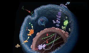
A paper published on October 2 in the Journal of Virology describes an exciting development in the world of influenza research—the construction of a luciferase reporter virus that does not affect virulence and can be used to track development and spread of infection in mice.

Insertion of luciferase reporter genes into viruses has been accomplished before for several viruses, but has not been successful for influenza. Construction of influenza reporter viruses is complicated because the viral genome is small and all the viral genes are critical for infection. Therefore, replacement of an existing gene with a reporter gene or insertion of additional reporter sequences without affecting the virus’s ability to replicate and cause infection has proven difficult. To be successful, a reporter gene needs to be small enough to insert into the viral genome without eliminating any other vital functionality.

Continue reading “NanoLuc® Luciferase: A Good Thing for Small Packages”

Piecing the Puzzle Together: Using Multiple Assays to Better Understand What Is Happening with Your Cells

You often need several pieces of information to really understand what is happening within a cell or population of cells. If your cells are not proliferating, are they dying? Or, are you seeing cytostasis? If they are dying, what is the mechanism? Is it apoptosis or necrosis? If you are seeing apoptosis, what is the pathway: intrinsic or extrinsic?

If you are measuring expression of a reporter gene and you see a decrease in expression, is that decrease due to transfection inefficiencies, cytotoxicity, or true down regulation of your reporter gene?

To investigate these multiple parameters, you can run assays in parallel, but that requires more sample, and sample isn’t always abundant.

Multiplexing assays allows you to obtain information about multiple parameters or events (e.g., reporter gene expression and cell viability; caspase-3 activity and cell viability) from a single sample. Multiplexing saves sample, saves time and gives you a more complete picture of the biology that is happening with your experimental sample.

What information do you need about your cells to complete the picture?

Multiplexing assay reagents to measure biomarkers in the same sample has often been considered an application only accomplished with antibodies or dyes and sophisticated detection instrumentation. However, Promega has developed microwell plate based assays for cells in culture that allow multiplexed detection of biomarkers in the same sample well using standard multimode multiwell plate readers. Continue reading “Piecing the Puzzle Together: Using Multiple Assays to Better Understand What Is Happening with Your Cells”

A Clean Brain Is a Healthy Brain

College student sleeping instead of studying
Can you sleep your way to a “cleaner” brain?
It is hard to undermine the role of cleanliness in disease prevention, both internally and externally. Within our body, the lymphatic system plays an important role in clearing the intercellular passages of large and potentially harmful toxic molecules and recirculate back into the blood stream. This enables the transport of these molecules to liver for inactivation and subsequent removal from the body. Therefore, lymphatic system prevents build-up of soluble proteins in the interstitial space. Typically, more metabolically active a cell is, more intricate is the lymphatic vasculature around it. This observation was in contrast to our scientific knowledge a few years ago, when we believed that due to the presence of the blood-brain barrier, there was no lymphatic system active in the brain. The brain, as we know, is highly active metabolically and the removal of harmful solutes and proteins from the neuronal vicinity is of utmost urgency. For a long time it was believed that cerebrospinal fluid (CSF), while coursing through the brain also removed cellular metabolite by products, apart from carrying nutrients to brain tissue, through a process known as diffusion. This is a rather slow process and it did not very well explain how large molecules such as proteins were removed from the interstitial place.

Recently, using two-photon imaging technique in live mice, scientists at Rochester discovered (1) that there is another vasculature functioning in the brain which circulates CSF to every corner of the brain much more efficiently, through bulk flow or convection. Continue reading “A Clean Brain Is a Healthy Brain”

Detecting Apoptosis

The concept of cell death as a normal cell fate was articulated only three years after Schleiden and Schwann introduced the Cell Theory when, in 1874, Vogt described natural cell death as an integral part of toad development (as cited in Cotter and Curtin, 2003). Since these early observations, natural cell death has been described “anew” several times. In 1885 Flemming provided the first morphological description of a natural cell death process, which we now label “apoptosis”, a term coined by Kerr and colleagues to describe the unique morphology associated with a cell death that differs from necrosis (as cited in Kerr et al. 1972).

Apoptosis morphology
Progression of morphology changes during apoptosis.

In the 1970s and 1980s, studies revealed that apoptosis not only had specific morphological characteristics but that it was also a tightly regulated process with specific biochemical characteristics. Studies of cell lineage in the nematode, Caenorhabditis elegans, showed that apoptosis was a normal feature of the nematode’s invariant developmental program (Hengartner, 1997). At the biochemical level, Wyllie showed that DNA degradation by a specific endonuclease during apoptosis resulted in a DNA ladder composed of mono- and oligonucleosomal-sized fragments (Wyllie, 1980).

These and many other studies have proven that apoptosis is a critical component of development, and when it doesn’t happen appropriately, it can be pathological, leading to cancers or other diseases. Therefore, understanding how and when apoptosis occurs and the many signals that can trigger this process is a focus of many laboratory experiments.

There are many ways to detect apoptosis in cells or tissues. This blog describes some of the most common ones.

Continue reading “Detecting Apoptosis”

Elevating “bliss” the natural way

Sun Runners
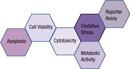
Marijuana is a highly controversial substance with roughly an equal number of supporters and opponents of its use for medicinal purposes. Marijuana is a dry, shredded mix of flowers, stems, seeds and leaves of the Hemp plant Cannabis sativa. New studies reporting the efficacy of medicinal marijuana in clinical conditions surface on a fairly regular basis, with the latest being a reported treatment for seizures. This constant influx of new information shows how little we know about the substance and how it works in the human body. So what do we know about this substance? While many psychoactive drugs clearly fall into the category of either stimulant, depressant or hallucinogen, Cannabis exhibits a mix of all properties, perhaps leaning the most towards hallucinogenic or psychedelic, though with other effects quite pronounced as well. Continue reading “Elevating “bliss” the natural way”

Shining a Bright Light on Deep Questions in Biology with Bioluminescence

artists view inside a cell

Search the PubMed database for “dual-luciferase” and citations abound. The Dual-Luciferase® Reporter Assay is a powerful tool that allows researchers to ask a multitude of questions about gene control and expression in a system that itself could be normalized and internally controlled. For more than 15 years, firefly and Renilla luciferases  have formed the basis of a range of powerful assays and research tools for scientists who are asking questions about the deep and complex genetic and cellular story associated with cancer. Here we talk a bit of about bioluminescent chemistries, some of the newest bioluminescent tools available, and how some of these tools can be used to probe the deeper questions of cell biology, including cancer biology.

Continue reading “Shining a Bright Light on Deep Questions in Biology with Bioluminescence”

Screening for Inhibitors of CD73 (5´-ectonucleotidase) Using a Metabolite Assay

CD73

CD73 also known as 5´-Ectonucleotidase (NT5E) is a membrane-anchored protein that acts at the outer surface of the cell to convert AMP to adenosine and free phosphate. CD73 activity is associated with immunosuppression and prometastatic effects, including angiogenesis. CD73 is highly expressed on the surfaces of many types of cancer cells and other immunosuppressive cells (1). A recent study by Quezada and colleagues showed that the high concentration of adenosine produced by the CD73-catalyzed reaction on glioblastoma multiforme cells, which are characterized by extreme chemoresistance, triggered adenosine signaling and in turn, the multi-drug resistance (MDR) phenotype of these cells (2).

Because of the roles of adenosine in immunosuppression, angiogenesis and MDR phenotypes, CD73 (NT5E) is an attractive therapeutic target. However, the current methods of assaying for the ectonucleotidase activity, HPLC and a malachite green assay, are cumbersome and not suited to high-throughput screening. The HPLC assay is expensive and difficult to automate and miniaturize (3). The malachite green assay is sensitive to phosphate found in media, buffers and other solutions used in the compound-screening environment.

To address the problem of developing a reliable high-throughput screening assay for CD73, Sachsenmeier and colleagues (3) looked to a luminescent ATP-detection reagent.

Continue reading “Screening for Inhibitors of CD73 (5´-ectonucleotidase) Using a Metabolite Assay”

Another Step Closer to Understanding Epigenetic Gene Regulation

Chromatin fiber

Back when I was a graduate student (more than a few years ago), I remember hearing another student joke that if a member of his thesis committee asked him to explain an unexpected or unusual result, he was going to “blame” epigenetics. At that time, the study of epigenetic gene regulation was in its infancy, and scientists had much to learn about this mysterious regulatory process. Fast forward to today, and you’ll find that scientists know a lot more about basic epigenetic mechanisms, although there is still plenty to learn as scientists discover that the topic is much more complicated than initially thought, as is often the case in science. A recent EMBO Journal article is contributing to our knowledge by shedding light on the role of the TET family of DNA-modifying enzymes in epigenetics (1).

Continue reading “Another Step Closer to Understanding Epigenetic Gene Regulation”

“Fingerprinting” Your Cell Lines

Working in the laboratoryResearchers working with immortalized cell lines would readily agree when I state that it is almost impossible to look at cells under the microscope and identify them by name. There are phenotypic traits, however they do change with change in media composition, passage number and in response to growth factors. I remember the pretty arborizations my neuroblastoma cell line SH-SY5Y exhibited in response to nerve growth factor treatment. Thus physical appearance is not a distinguishing feature. Currently, in many labs, researchers typically use more than one cell line, and more than likely, share the same lab space to passage cells and the same incubator to grow the cells. In such scenarios, it is not difficult to imagine that cell lines might get mislabeled or cross-contaminated. For example HeLa cells, one of the fastest growing cell lines have been shown to invade and overtake other cell lines.

Misidentification of cell lines has deep and severe implications. A review of cell lines used to study esophageal adenocarcinoma found that a large number of the cell lines were actually derived from lung or gastric cancers. Unfortunately, by the time this error was discovered, data from these cell line studies were already being used for clinical trials and other advanced studies and publications. Moreover, the cell lines were being to screen and design and test specific cancer drugs which ended up in flawed clinical trials. Continue reading ““Fingerprinting” Your Cell Lines”

Chromatography and “Air Traffic Control” Interplay Direct Olfactory Function

It is not difficult to appreciate why a keen sense of smell is important to well-being and to general living. While it signals the presence of delicious (or stale) food before we can even see or taste it, it has obvious great survival value to be able to alert living beings of danger such as certain poisons, leaking gas or fire. Humans are known to identify about 10,000 different types of odors. Of course dogs have vastly improved and keener sense of smell than human beings.

When odorant molecules (molecules that we can smell) reach the nostril, they dissolve in the mucus and bind to olfactory receptors present on the cilia of each sensory neuron. This binding activates a G-protein coupled cascade involving adenylyl cyclase. This causes the release of cyclic AMP and opening of cAMP-dependent sodium channels. Influx of sodium causes the membrane to depolarize and activate an action potential for propagation of the signal to the brain where it is analyzed and decoded(1). This seems pretty straightforward until one realizes the sheer magnitude of smells we are able to identify using this mechanism. Continue reading “Chromatography and “Air Traffic Control” Interplay Direct Olfactory Function”