RNase, back in the early 1990s, posed a serious threat to laboratories working with RNA isolation. My graduate work involved isolating RNA from the tissues of Lyme disease-infected mice and hamsters. We struggled to DEPC-treat glass and plasticware, or autoclave anything that could be autoclaved, kept tissues cold during RNA harvest and held our breaths (truly, as aerosol could be another source of ribonuclease) until PAGE proved us successful in RNA isolation.
Ribonuclease (RNase) was omnipresent and the arch rival of our work, across several species, due to its RNA destroying abilities.
Now, a July 13, 2015 publication by researchers at the University of Wisconsin-Madison provided both a catch-up for this former lab rat on modern day research with and knowledge of RNase, as well as an exciting look at what may be a real purpose for this RNA-destroying molecule: RNase has moved to clinical trials due to the discovery of it’s cytotoxicity for cancer cells.
Raines’ group in the Department of Chemistry at UWI-Madison published in ACS Central Science their findings on the ligand that RNase 1 uses to attach to human cancer cells, in the article, “Human Cancer Antigen Globo H is a Cell-Surface Ligand for Human Ribonuclease 1”.
About Ribonucleases
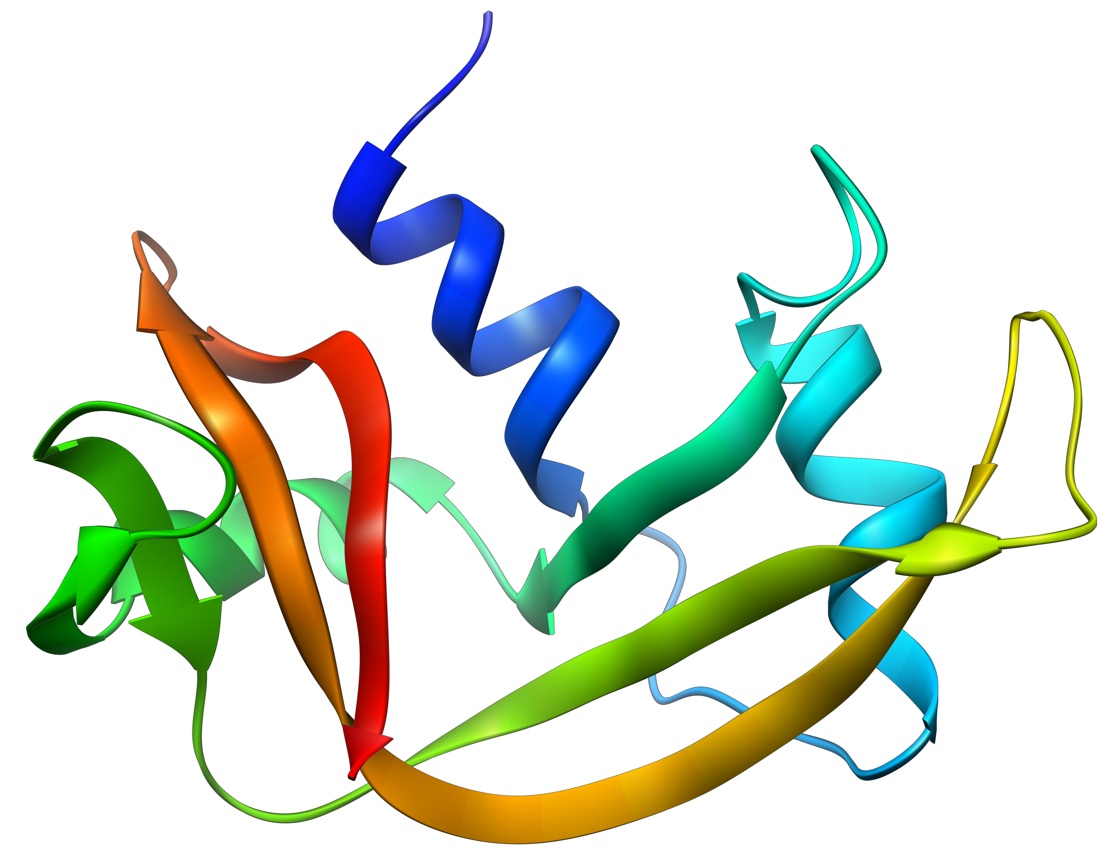
You may have tried to isolate RNA from organ tissues (i.e., liver, pancreas, kidney) and recall that RNase levels from certain organs were much higher than those found in other tissues. In fact the pancreas is a major source of RNase for vertebrates. RNase A is a well-studied enzyme from cows, with RNase 1 the most prevalent human cohort to RNase A. Both enzymes are highly efficient at chewing up RNA.
In previous work, Raines’ group engineered both enzymes to develop RNases that could evade a cytosolic ribonuclease inhibitor protein (1), resulting in RNases with cytotoxic abilities.
Mechanism of Cytotoxicity
Further work by this group showed a probable mechanism for the cell toxicity of these engineered RNases to be internalization of the RNase through endosomes, and translocation to the cytosol resulting in cleavage of RNA and apoptosis. of the cell.
Unexpected Finding
A truly exciting finding about these cytotoxic, engineered RNases was their specific targeting of cancer cells. In fact, a variant of RNase 1 is currently in clinical trials as a cancer chemotherapeutic agent. In this work they sought a specific RNase receptor on cancer cells.
Several Important Backstories
In earlier work it was shown that the surface of cancer cells has a greater anionic (negative) charge than that of noncancerous cells. In addition and perhaps due to their nature as rapidly dividing cells, cancer cells undergo endocytosis more rapidly than comparable noncancerous cells. While contributing, this data could explain only in part, the preferential uptake of RNase by cancer cells.
Previous work had also determined that one of an extensive series of polysaccharide-containing receptors on the surface of eukaryotic cells, is a glycan called Globo H. Globo H is a component of the outer membrane of epithelial cells from tissue sources like mammary, uterine, pancreas and kidney. It is noteworthy that Globo H is identified in far higher amounts on the outside of tumor specimens from lung, breast, prostate, pancreas, ovarian and other tumors, and histopathology has correlated the presence of high levels of Globo H on tumors with poor cancer outcomes.
It has been speculated that Globo H on the surface might help cancer cells evade immune detection. And once internalized, Globo H binding to translin-associated factor X has been shown to promote angiogenesis (1). Not surprisingly, vaccines based on Globo H are currently in clinical trials. Even so, not much is known about a functional role for Globo H.
In this Report
Using a printed array of eukaryotic cell surface glycans, the authors of this report showed that RNase A binds to Globo H. They next measured the affinity of bovine RNase A and its human cohort, RNase 1, for Globo H, and found RNase 1 to have greater affinity to Globo H. Through the use of antagonists, they showed that breast adenocarcinoma cells with lower amounts of surface Globo H, were less susceptible to cytotoxic RNases.
Finally, these authors demonstrated a connection between two endogenous cellular molecules that are currently and independently in clinical trials as cancer therapeutics, Globo H and RNase 1. In addition, Raines et al. speculate that the affinity of RNase 1 for Globo H may have evolved as an innate defense against cancer.
Although the most exciting news here is anticipating the good news that clinical trials may bring to cancer patients, there is also some satisfaction in knowing that all of that RNase we worked so hard to avoid has a purpose in host defense against a serious and in many cases still, incurable disease.
Reference
- Eller, C.H. et al. (2015) Human Cancer Antigen Globo H Is a Cell-Surface Ligand for Human Ribonuclease 1. ACS Central Science.
DOI: 10.1021/acscentsci.5b00164
Kari Kenefick
Latest posts by Kari Kenefick (see all)
- What Drives Muscle Fiber Shifts in Obesity and Type ll Diabetes? - October 9, 2025
- How Thalidomide and Molecular Glues Are Redefining Drug Discovery - August 21, 2025
- ATP-Powered Proteins Beyond Kinases – and Why Helicases Are Stealing the Spotlight - August 7, 2025