SARS-CoV-2 Nucleocapsid Protein and PA28γ: A Role in Pathogenesis?

The SARS-CoV-2 nucleocapsid protein accounts for the largest proportion of viral structural proteins and is the most abundant protein in infected cells. Nucleocapsid proteins have the job of “packaging” the viral nucleic acid (in this case, RNA). Viral nucleocapsid proteins can also enter the host nucleus and interact with a variety of host proteins to interfere with critical processes of the host cell, including protein degradation. Here we review a study that used an in vitro protein degradation assay to investigate the interaction of the SARS-CoV-2 nucleocapsid protein and the proteasome activator PA28γ.
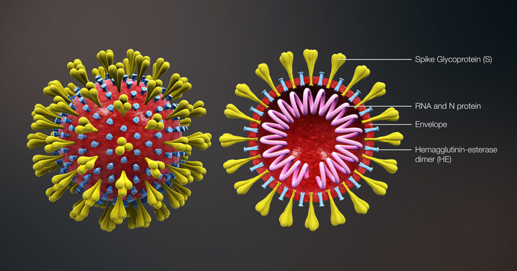
SARS-CoV-2 structural diagram, showing the SARS-CoV-2 nucleocapsid protein composed of RNA and N protein.

In SARS-CoV-2 infections, the nucleocapsid protein is critical for infection, replication and packaging. The SARS-CoV-2 nucleocapsid protein is not only localized in the cytosol of the host cell but also is translocated into the nucleus. There, it interacts with various cellular proteins that modulate cellular functions, such as the degradation of unneeded or damaged proteins by proteolysis. Researchers have proposed that the protein degradation system plays an important part in coronavirus infection (1).

Continue reading “SARS-CoV-2 Nucleocapsid Protein and PA28γ: A Role in Pathogenesis?”
Go fISH! Using in situ Hybridization to Search for Expression of a SARS-CoV-2 Viral Entry Protein

Loss of smell (olfaction) is a commonly reported symptom of COVID-19 infection. Recently, Bilinska, et al. set out to better understand the underlying mechanisms for loss of smell resulting from SARS-CoV-2 infection. In their research, they used in situ hybridization to investigate the expression of TMPRSS2, a SARS-CoV-2 viral entry protein in olfactory epithelium tissues of mice.

Continue reading “Go fISH! Using in situ Hybridization to Search for Expression of a SARS-CoV-2 Viral Entry Protein”

NanoBRET™ Assays to Analyze Virus:Host Protein:Protein Interactions in Detail

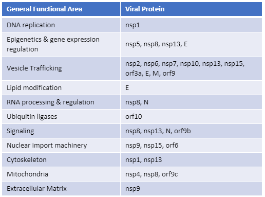
Recently, Gordon et al. published an atlas of protein:protein interactions of all proposed SARS-CoV-2 proteins expressed individually in HEK 293 cells (Table 1). The study tagged each of the viral proteins with an epitope tag and performed a pull-down of the expressed protein followed by trypsin digestion and mass spec analysis, a process referred to as affinity purification–mass spec analysis. The group identified 332 human proteins interacting with 27 SARS-CoV-2 proteins.

The interactions identified in the HEK 293 cells helped Appelberg et al. analyze interactions over time in SARS-CoV-2-infected Huh7 cells. Gordon et al. used the PPI data to identify FDA-approved drugs, drugs in clinical trials, and pre-clinical compounds that bound to the identified human proteins and labs in New York and Paris tested some of these drugs for antiviral effects.   

Table 1. The general functional area of human proteins identified to interact with individually expressed SARS-CoV-2 proteins as reported by Gordon, et al. (1). The SARS-CoV-2 proteins are classified as non-structural proteins (nsp#), structural proteins (E, M, and N) and accessory proteins (orf#).  
Continue reading “NanoBRET™ Assays to Analyze Virus:Host Protein:Protein Interactions in Detail”