Elegant Experiments that Changed the World

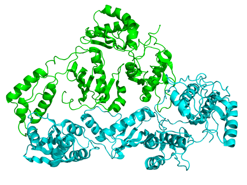
Crystallographic structure of HIV reversed transcriptase. Wikimedia Commons
Crystallographic structure of HIV reverse transcriptase. Wikimedia Commons

Today, reverse transcriptases are commonplace molecular biology tools, easy to obtain and routinely used in labs for everyday cloning and gene expression analysis experiments. Reverse transcriptase inhibitors have also found widespread use as antiviral drugs in the treatment of retroviral infections.

It’s easy to forget that the existence of reverse transcriptase activity—the ability to convert an RNA template into DNA—was once a revolutionary notion not easily accepted by the scientific community. The idea that RNA could be the template for DNA synthesis challenged the “DNA–>RNA–> Protein” central dogma of molecular biology.

The foundational studies that proved the existence of a reverse transcriptase activity in RNA tumor viruses were described in two papers published back-to-back in Nature in June, 1970. Two of the authors of these studies, Howard Temin of the University of Wisconsin and David Baltimore of the Massachusetts Institute of Technology, were awarded a Nobel Prize for their work in 1975.

In appreciation of the significance of these papers, the editorial introduction published in Nature at the time states:

This discovery, if upheld, will have important implications not only for carcinogenesis by RNA viruses but also for the general understanding of genetic transcription: apparently the classical process of information transfer from DNA to RNA can be inverted.

Before these papers were published, it was known that successful infection of cells by RNA tumor viruses required DNA synthesis. Formation of virions could be inhibited by Actinomycin D—an inhibitor of DNA-dependent RNA polymerase—so it was known that synthesis of viral RNA from a DNA template was part of the viral life cycle. The existence of an intracellular DNA viral genome was therefore indicated, and had been postulated by Temin in the mid 1960’s. However, proof of the mechanism whereby this DNA template was generated from the RNA genome of the infecting virus remained elusive. Continue reading “Elegant Experiments that Changed the World”
ProK: An Old ‘Pro’ That is Still In The Game

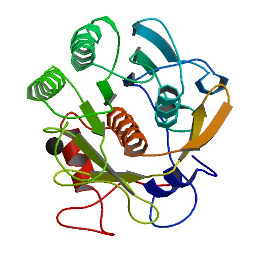
Proteinase K Ribbon Structure ImageSource=RCSB PDB; StructureID=4b5l; DOI=http://dx.doi.org/10.2210/pdb4b5l/pdb;

If you enter any molecular lab asking to borrow some Proteinase K, lab members are likely to answer: “I know we have it. Let me see where it is”. Sometimes the enzyme will be found to have expired. The lab may also have struggled with power outages or freezer malfunctions in the past. But the lab still decides to keep the enzyme. One may rightly ask – why do labs hang on to Proteinase K even when it has been stored under sub-standard conditions?

Continue reading “ProK: An Old ‘Pro’ That is Still In The Game”

DNA Purification, Quantitation and Analysis Explained

WebinarsYesterday I listened in on the Webinar “Getting the Most Out of Your DNA Analysis from Purification to Downstream Assays”, presented by Eric Vincent–a Product Manager in the Promega Genomics group.

This is the webinar for you if you have ever wondered about the relative advantages and disadvantages of the many methods available for DNA purification, quantitation and analysis, or if you are comparing options for low- to high-throughput DNA purification. Eric presents a clear analyses of each of the steps in a basic DNA workflow: Purification, Quantitation, Quality Determination, and Downstream Analysis, providing key considerations and detailing the potential limitations of the methods commonly used at each step.

The DNA purification method chosen has an affect on the quality and integrity of the DNA isolated, and can therefore affect performance in downstream assays. Accuracy of quantitation also affects success, and the various downstream assays themselves (such as end-point PCR, qPCR, and sequencing) each have different sensitivities to factors such as DNA yield, quality, and integrity, and the presence of inhibitors. Continue reading “DNA Purification, Quantitation and Analysis Explained”

Cell free application: Sumoylation characterization

Small Ubiquitin-like Modifier (or SUMO) proteins are a family of small proteins that are covalently attached to and detached from other proteins in cells to modify their function. SUMOylation is a post-translational modification involved in various cellular processes, such as nuclear-cytosolic transport,
transcriptional regulation, apoptosis, protein stability and response to stress.

In contrast to ubiquitin, SUMO is not used to tag proteins for degradation. Mature SUMO is produced when the last four amino acids of the C-terminus have been cleaved off to allow formation of an isopeptide bond between the C-terminal glycine residue of SUMO and an acceptor lysine on the target protein.

Cell free expression can be used to characterize sumoylation of proteins. Target proteins are expressed in a rabbit reticulocyte cell free system (supplemented with necessary addition components,). Proteins that have been modified can be analyzed by a shift in migration on polyacrylamide gels, when compared to control reactions.

The following references illustrate the use of cell free expression for this application.

Brandl, A. et al. (2012) Dynamically regulated sumoylation of HDAC2 controls p53 deacetylation and restricts apoptosis following genotoxic stress. J. Mol. Cell. Biol. (online only)

Janer, A. et al. (2010). SUMOylation attenuates the aggregation propensity and cellular toxicity of the polyglutamine expanded ataxin-7. Human. Mol. Gen. 19, 181—95.

Rytinki, M. et al. (2009) SUMOylation attenuates the function of PGC-1alpha. J. Biol. Chem. 284, 26184-93.

Klein, U. et al. (2009) RanBP2 and SENP3 function in a mitotic SUMO2/3 conjugation-deconjugation cycle on Borealin. Mol. Cell. Biol. 20, 410–18.

Seo, W. and Ziltener, H. (2009) CD43 processing and nuclear translocation of CD43 cytoplasmic tail are required for cell homeostasis. Blood, 114, 3567–77.

A New Method that Marks Proteins for Destruction

The ability to manipulate genes and proteins and observe the effects of specific changes is a foundational aspect of molecular biology. From the first site-directed mutagenesis systems to the development of knockout mice and RNA interference, technologies for making targeted changes to specific proteins to eliminate their expression or alter their function have made tremendous contributions to scientific discovery.
A recent paper highlights a novel application of HaloTag technology to enable the targeted destruction of specific HaloTag fusion proteins in vivo. The paper, published online in the July issue of Nature Chemical Biology, details a promising new method with application for validation of potential drug targets by specific in vivo inhibition, and for studying the function of specific genes in organogenesis or disease development. Continue reading “A New Method that Marks Proteins for Destruction”

Antibody Art

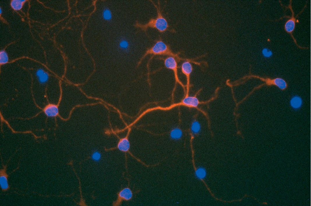
Double fluorescent staining of CNS cells with Anti-Rat ciliary neurotrophic factor. Nuclei stained with DAPI

There are many levels on which science can be a beautiful thing. Some of these are quite abstract, like the experimental result that exactly proves the theory, the perfect order revealed in a mathematical equation, or the exquisite sensitivity seen in regulation of a signaling pathway. On another level the output of an experiment itself can have a physical beauty of its own. For example, immunofluorescence and immunocytochemistry technologies can generate results that not only reveal information about the subject of the experiment, but also can be quite spectacularly beautiful. Here are a few favorites of mine:

Continue reading “Antibody Art”

Quorum Sensing in Bacteria: How a Picture can be Worth a Thousand Words

Increasingly, multimedia and video are being used in addition to traditional delivery methods to communicate scientific findings. Journals such as PLoS ONE, Cell, Nature and others often use video to either showcase particular articles, or offer authors the opportunity to include multimedia elements as part of their article. Some subjects lend themselves better to video delivery than others. Every so often a video report comes along that perfectly complements the content of the associated paper, illustrating the power of video to enhance communication of research findings.

In my opinion, the effective use of video to highlight results is beautifully illustrated by the report below, highlighting the publication “A synchronized quorum of genetic clocks” by Danino et al, which was published in Nature this week.

[youtube=http://www.youtube.com/watch?v=pnjdAr4EjI0]
Continue reading “Quorum Sensing in Bacteria: How a Picture can be Worth a Thousand Words”