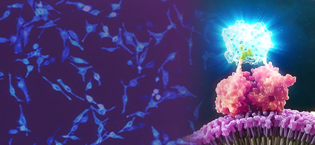
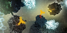
In biologics, cell therapy, and targeted protein degradation, the science is moving fast—and so are the tools. From GPCR-targeted therapies to real-time CAR-T manufacturing tools, new techniques are reshaping how scientists approach drug development, live-cell imaging, and protein degradation.
The “Bringing Light to Science”Discover Glo 2025 speaker series brought together researchers from across academia and industry to share real-world examples of how bioluminescent technologies are helping them advance their research. Now available on demand, these sessions offer fresh perspectives and actionable takeaways on the future of therapeutic development, cellular analysis and assay design.
We’ve distilled five key takeaways from the sessions—practical insights you can apply to your own work or use to stay current with where the field is heading.
Luminescent live-cell assays are powerful tools for cellular biology research. They offer both qualitative and quantitative insights into processes such as gene expression, cell viability, metabolic activity, protein and small molecule interactions, and targeted protein degradation. But what if you could go beyond the numbers and actually see what is happening in your cells? With luminescent imaging, you have the opportunity to uncover more dynamic data by visualizing what happens with your cells in real time.
Why Luminescent Imaging?
Bioluminescent reporters such as NanoLuc® luciferase are well-suited for bioluminescent imaging. NanoLuc, the smallest engineered luciferase available, is up to 150X brighter than firefly or Renilla luciferase, so exposure times can be shorter than they would be with other luminescent reporters. Its small size also makes it less likely to perturb normal biology or protein function.
Another benefit of bioluminescence for imaging is the inherent stability and sustainability of the bioluminescent signal, which does not require external excitation like fluorescent tags. This allows direct visualization of protein dynamics in living cells without the need for repeated sample excitation. The lack of external excitation also reduces the risk of phototoxicity and photobleaching, common issues that can adversely affect cell viability and signal integrity over time.
Applications Across Cellular Research
Luminescent imaging complements traditional luminescence assays by adding spatial and temporal dimensions. With luminescent live-cell imaging, researchers can visualize NanoLuc® Luciferase assays to gain a deeper understanding of the real-time cellular processes occurring in each experiment. Applications include:
Determining which cells provide signal
Analyzing mixed cell populations
Identifying rare events
Monitoring protein:protein interactions
Identifying protein localization and translocation
Tracking protein degradation and stability over time
Selectively targeting proteins for removal from the cell—instead of inhibiting protein activity—is a newer approach with therapeutic potential. In this method, the protein is targeted for degradation using the cell’s natural ubiquitin proteasome system (UPS). The degradation process is initiated by compounds such as molecular glues and proteolysis targeting chimeras (PROTACs) linking the target protein to an E3 ligase. Once this linkage occurs, the cell’s UPS does the rest.
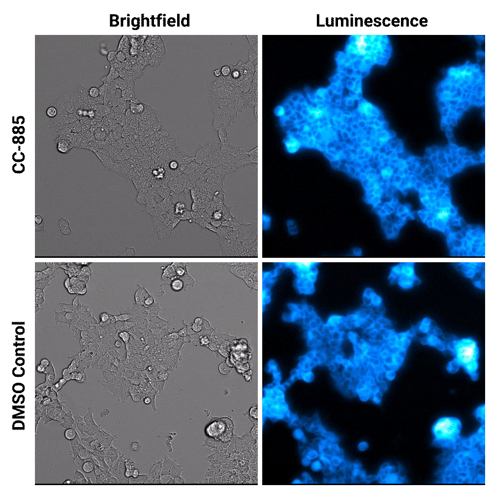
Luminescent substrates with increased signal stability, such as the Nano-Glo® Extended Live Cell Substrate, enable researchers to image targeted protein degradation in their cells in real time. In the example shown below, Nano-Glo® Vivazine™ Live Cell Substrate was used to image degradation of the GSPT1 protein by the CC-885 degrader over 5 hours.
Targeted protein degradation over time. HEK293 cells expressing endogenous HiBiT-tagged GSPT1 and stably expressing LgBiT were treated with CC-885 degrader or DMSO control treatment. Assayed with Nano-Glo® Vivazine™ Live Cell Substrate and imaged over 5 hours using GloMax® Galaxy Bioluminescence Imager.
Combining Luminescent and Fluorescent Imaging to Detect Protein:Small Molecule Interactions
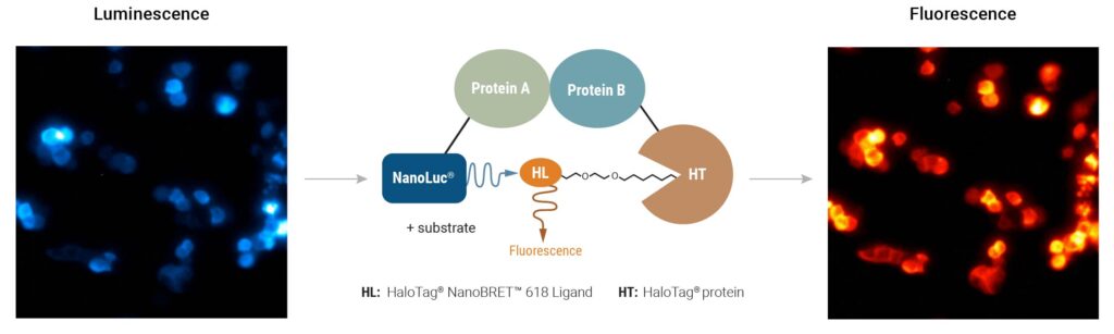
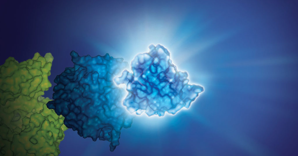
Using bioluminescence resonance energy transfer (BRET)-based assays such as NanoBRET® assays allows you to detect protein:protein interactions by measuring energy transfer from a bioluminescent protein donor to a fluorescent protein acceptor. These assays can be used to monitor changes in protein interactions over time, making them a useful tool for small-molecule screening.
The schematic below illustrates how the NanoBRET® NanoGlo® Detection Systems can be used to visualize target engagement. The cells on the left are expressing a NanoLuc® fusion protein, resulting in a luminescent signal. Adding a fluorescent small tracer (center) results in energy transfer and a fluorescent signal (right). Using an imaging platform that has luminescence and fluorescence imaging capabilities will let you see this energy transfer in action.
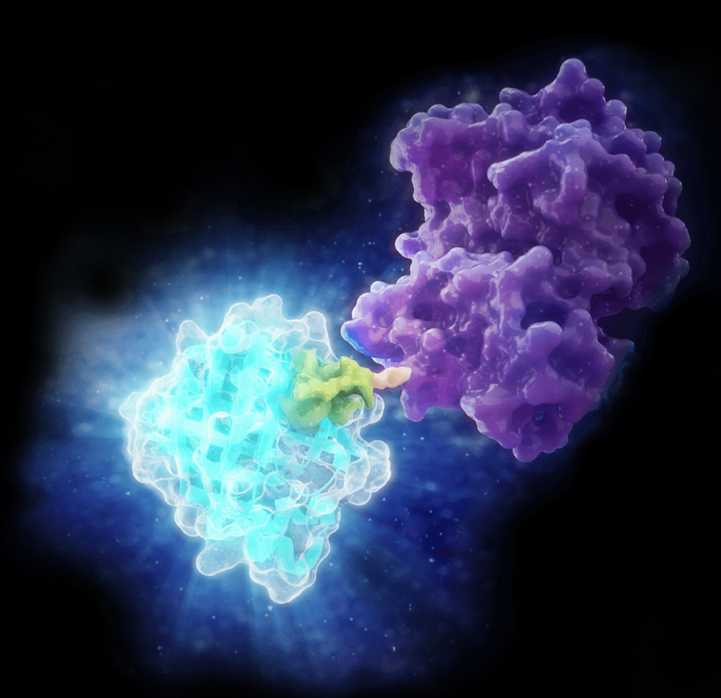
Detecting protein:small molecule interactions with NanoBRET® NanoGlo® Detection Systems. HCT116 cells expressing a PRMT5–NanoLuc® fusion were supplemented with a fluorescent small molecule tracer (center panel). Before tracer addition, luminescent signal indicates energy is present on the donor protein (left; 3-minute exposures for 15 minutes). Binding of fluorescent tracer results in energy transfer and fluorescent signal (right; 3-minute exposures for 60 minutes). Images were captured on the GloMax® Galaxy Bioluminescence Imager.
Bringing the Power of Luminescent Imaging to Your Lab
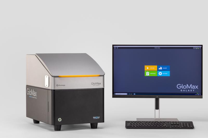
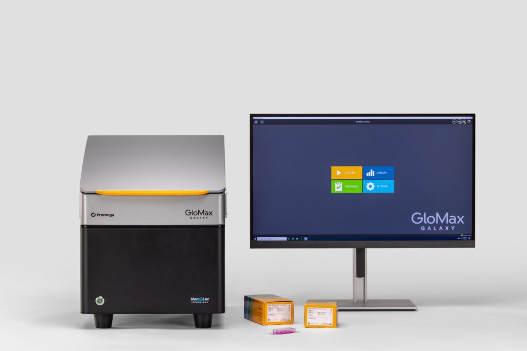
The GloMax® Galaxy Bioluminescence Imager was built to visualize the NanoLuc®-based assays we engineered, in a benchtop instrument accessible to any lab. The Galaxy is a fully equipped microscope with luminescence, fluorescence, and brightfield imaging capabilities, giving researchers a way to see functional and dynamic cellular events across a cell population.
Conclusion
Luminescent imaging can enrich what we learn from live-cell assays and offers an unprecedented view into the dynamics of cellular processes. From monitoring drug responses to visualizing protein interactions, this technology delivers insights that go beyond the capabilities of traditional assays.
Whether you’re studying cancer biology, drug development or cellular signaling, luminescent imaging can help you uncover what’s hidden in your data and see your research in a whole new light.
GloMax® Galaxy Luminescent Imager, NanoBRET® Nano-Glo® Detection Systems and Nano-Glo® Vivazine live Cell Substrate are for Research Use Only. Not for Use in Diagnostic Procedures.
If you’re familiar with bioluminescence, you’ve probably used it in plate-based experiments to track various biological processes. You understand it provides distinct advantages over traditional fluorescence assays, particularly when it comes to sensitivity. However, there’s always that one nagging question: how representative is the signal on a cell-to-cell level?
Traditional approaches to decipher cell-to-cell signal rely on complex, time-intensive measures that only approximated the findings acquired through bioluminescence. That’s where the GloMax® Galaxy Bioluminescence Imager comes in. Built to visualize the NanoLuc®-based assays we engineered, the Galaxy goes beyond simple numeric outputs to reveal what’s happening in individual cells.
NanoLuc® luciferase is up to 150X brighter than firefly or Renilla luciferase, giving it the sensitivity to detect subtle protein interactions in complex biological systems. This bright luminescent enzyme enables a much broader linear range than fluorescence, improving detection of small changes in protein activity, such as proteins interacting. Microplate readers measuring NanoLuc® assays rely on signal generated from many cells. This results in an approximation of what is occurring biologically. Truly validating those luminescent readings within a cell population has been challenging—until now. The GloMax® Galaxy allows you to perform bioluminescence imaging, moving beyond the numbers, offering the power to visualize protein interactions directly.
Recombinant adeno-associated viral (AAV) vectors are an appealing delivery strategy for in vivo gene therapy but face a formidable challenge: avoiding detection by an ever-watchful immune system (1,2). Efforts to compensate for the immune response to these virus particles have included immunosuppressive drugs and engineering the AAV vector to be especially potent to minimize its effective dosage. These methods, however, come with their own challenges and do not directly solve for the propensity of AAV vectors to induce immune responses.
A recent study introduced a new approach to reduce the inherent immunogenicity of AAV vectors (2). Researchers strategically swapped out amino acids in the AAV capsid to remove the specific sequences recognized by T-cells that elicit the most pronounced immune response. As a result, they significantly reduced T-cell mediated immunogenicity and toxicity of the AAV vector without compromising its performance.
With advancements made over the past few decades, the future of in vivo bioluminescence imaging (BLI) continues to gain momentum. In vivo BLI provides a non-invasive way to image endogenous biological processes in whole animals. This provides an easier method to assess relevant systems and functions. Unlike fluorescent imaging, BLI relies on a combination of enzymes and substrates to produce light, greatly reducing background signal (Refaat et al., 2022). Traditional fluorescent tags are also quite large and may interfere with normal biological function. In vivo BLI research has been around for quite some time, primarily utilizing Firefly luciferase (Luc2/luciferin). A recent advancement was the creation of the small and bright NanoLuc® luciferase (NLuc). Promega offers an wide portfolio of NLuc products that provide ways to study genes, protein dynamics, and protein:protein interactions. To fully grasp the power of these tools, I interviewed several key investigators to determine their perspectives on the future of in vivo BLI. I was specifically interested in their thoughts on NLuc multiplexing potential with Firefly (FLuc), and future research areas. These two investigators are Dr. Thomas Kirkland, Sr. Scientific Investigator at Promega, and Dr. Laura Mezzanotte, Associate Professor at Erasmus MC.
Bioluminescence imaging is a powerful tool for non-invasive studies of the effect of treatments on cells and tissues. The luminescent signal is strong, and can be used in vivo, enabling repeated observations over time, allowing longitudinal study of cellular changes for hours or days. Bioluminescence imaging can be used in live animals over varying periods of time, without interfering with normal cellular processes.
Fluorescence imaging is also used in cellular studies. Although it can provide a stronger signal than luminescence, fluorescence requires light for excitation, and thus its in vivo use is limited at a tissue or cell depth greater than 1mm.
NanoLuc® Luciferase. Small, bright and now useful in brain bioluminescence imaging.
In addition, autofluorescence can be an issue with fluorescence imaging, as cellular components and surrounding proteins and cells can fluoresce when exposed to light. Autofluorescence can result in high background signals, making it difficult to distinguish true fluorescence from background.
Many deep sea creatures are bioluminescent. However, before documenting the luminescence of the kitefin shark, Dalatias licha, there has never been a nearly six-foot long luminous vertebrate creature. In a recent study, Mallefet and colleagues examined three species of sharks: Dalatias licha, Etmopterous lucifer, and Emopterus granulosus and documented their luminescence for the first time. These bioluminescent sharks are the largest bioluminescent creatures known.
Studying protein function in live cells is limited by the tools available to analyze the expression and interactions of those proteins. Although mass spectrometry and antibody-based protein detection are valuable technologies for protein analysis, both methods have drawbacks that limit the range of targets and contexts in which proteins can be investigated.
Mass spectrometry is often poor at detecting low-abundance proteins. Antibody-based techniques require high quality, specific antibodies, which can be difficult to impossible to acquire. Both methods require cell lysis, preventing real-time analysis and limiting the physiological relevance, and both methods can be limiting for higher-throughput analysis. While plasmid-based overexpression of tagged target proteins simplifies detection and can allow for real time analysis, protein levels don’t typically resemble endogenous levels. Overexpression also has the potential to create experimental artifacts or limit the dynamic range of an observed response.
In 2018, Promega R&D scientists published a paper in ACS Chemical Biology demonstrating the use of CRISPR/Cas9 to integrate the 11 amino acid, bioluminescent HiBiT tag directly into the genome to serve as an easily measured reporter for endogenous proteins. This provides a highly quantitative method for investigating cellular protein dynamics that sidesteps the need for cloning and other drawbacks to conventional methods, including the ability to measure changing protein dynamics in real-time. (For more details about CRISPR/Cas9 knock-in tagging and other applications, read this blog.)
While their findings showed that this method provides efficient and specific tagging of endogenous proteins, the research was limited to just five different proteins within a single signaling pathway in two cell lines. This left unanswered questions about whether this approach was scalable, had broader applications and how accurately the natural biology of the cells was represented.
Live animal in vivo imaging is a common and useful tool for research, but current tools could be better. Two recent papers discuss adaptations of BRET technology combining the brightness of fluorescence with the low background of a bioluminescence reaction to create enhanced in vivo imaging capabilities.
The key is to image photons at wavelengths above 600nm, as lower wavelengths are absorbed by heme-containing proteins (Chu, J., et al., 2016 ). Fluorescent protein use in vivo is limited because the proteins must be excited by an external light source, which generates autofluorescence and has limited penetration due to absorption by tissues. Bioluminescence imaging continues to be a solution, especially firefly luciferase (612nm emission at 37°C), but its use typically requires long image acquisition times. Other luciferases, like NanoLuc, Renilla, and Gaussia, etc. either do not produce enough light or the wavelengths are readily absorbed by tissues, limiting their use to near-surface imaging.
The two papers discussed here illustrate how researchers have combined NanoLuc® luciferase with a fluorescent protein to harness bioluminescent resonance energy transfer (BRET) for brighter in vivo imaging reporters.
XWe use cookies and similar technologies to make our website work, run analytics, improve our website, and show you personalized content and advertising. Some of these cookies are essential for our website to work. For others, we won’t set them unless you accept them. To learn more about our approach to Privacy we invite you to Read More
By clicking “Accept All”, you consent to the use of ALL the cookies. However you may visit Cookie Settings to provide a controlled consent.
We use cookies and similar technologies to make our website work, run analytics, improve our website, and show you personalized content and advertising. Some of these cookies are essential for our website to work. For others, we won’t set them unless you accept them. To find out more about cookies and how to manage cookies, read our Cookie Policy.
If you are located in the EEA, the United Kingdom, or Switzerland, you can change your settings at any time by clicking Manage Cookie Consent in the footer of our website.
Necessary cookies are absolutely essential for the website to function properly. These cookies ensure basic functionalities and security features of the website, anonymously.
Cookie
Duration
Description
cookielawinfo-checbox-analytics
11 months
This cookie is set by GDPR Cookie Consent plugin. The cookie is used to store the user consent for the cookies in the category "Analytics".
cookielawinfo-checbox-functional
11 months
The cookie is set by GDPR cookie consent to record the user consent for the cookies in the category "Functional".
cookielawinfo-checbox-others
11 months
This cookie is set by GDPR Cookie Consent plugin. The cookie is used to store the user consent for the cookies in the category "Other.
cookielawinfo-checkbox-advertisement
1 year
The cookie is set by GDPR cookie consent to record the user consent for the cookies in the category "Advertisement".
cookielawinfo-checkbox-necessary
11 months
This cookie is set by GDPR Cookie Consent plugin. The cookies is used to store the user consent for the cookies in the category "Necessary".
cookielawinfo-checkbox-performance
11 months
This cookie is set by GDPR Cookie Consent plugin. The cookie is used to store the user consent for the cookies in the category "Performance".
gdpr_status
6 months 2 days
This cookie is set by the provider Media.net. This cookie is used to check the status whether the user has accepted the cookie consent box. It also helps in not showing the cookie consent box upon re-entry to the website.
lang
This cookie is used to store the language preferences of a user to serve up content in that stored language the next time user visit the website.
viewed_cookie_policy
11 months
The cookie is set by the GDPR Cookie Consent plugin and is used to store whether or not user has consented to the use of cookies. It does not store any personal data.
Analytical cookies are used to understand how visitors interact with the website. These cookies help provide information on metrics the number of visitors, bounce rate, traffic source, etc.
Cookie
Duration
Description
SC_ANALYTICS_GLOBAL_COOKIE
10 years
This cookie is associated with Sitecore content and personalization. This cookie is used to identify the repeat visit from a single user. Sitecore will send a persistent session cookie to the web client.
vuid
2 years
This domain of this cookie is owned by Vimeo. This cookie is used by vimeo to collect tracking information. It sets a unique ID to embed videos to the website.
WMF-Last-Access
1 month 18 hours 24 minutes
This cookie is used to calculate unique devices accessing the website.
_ga
2 years
This cookie is installed by Google Analytics. The cookie is used to calculate visitor, session, campaign data and keep track of site usage for the site's analytics report. The cookies store information anonymously and assign a randomly generated number to identify unique visitors.
_gid
1 day
This cookie is installed by Google Analytics. The cookie is used to store information of how visitors use a website and helps in creating an analytics report of how the website is doing. The data collected including the number visitors, the source where they have come from, and the pages visted in an anonymous form.
Advertisement cookies are used to provide visitors with relevant ads and marketing campaigns. These cookies track visitors across websites and collect information to provide customized ads.
Cookie
Duration
Description
IDE
1 year 24 days
Used by Google DoubleClick and stores information about how the user uses the website and any other advertisement before visiting the website. This is used to present users with ads that are relevant to them according to the user profile.
test_cookie
15 minutes
This cookie is set by doubleclick.net. The purpose of the cookie is to determine if the user's browser supports cookies.
VISITOR_INFO1_LIVE
5 months 27 days
This cookie is set by Youtube. Used to track the information of the embedded YouTube videos on a website.
Performance cookies are used to understand and analyze the key performance indexes of the website which helps in delivering a better user experience for the visitors.
Cookie
Duration
Description
YSC
session
This cookies is set by Youtube and is used to track the views of embedded videos.
_gat_UA-62336821-1
1 minute
This is a pattern type cookie set by Google Analytics, where the pattern element on the name contains the unique identity number of the account or website it relates to. It appears to be a variation of the _gat cookie which is used to limit the amount of data recorded by Google on high traffic volume websites.