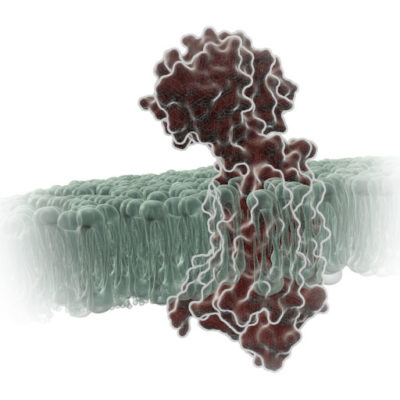
G Protein-Coupled Receptors (GPCRs) are a very large, diverse family of transmembrane receptors in eukaryotes. These receptors detect molecules outside the cell and activate internal signaling pathways by coupling with G proteins. Once a GPCR is activated, β-arrestins translocate to the cell membrane and bind to the occupied receptor, uncoupling it from G proteins and promoting its internalization.
Reporter tags are useful for studying the dynamics of GPCRs and associated proteins, but large tags can disrupt the receptors’ native functioning, and often overexpression of the tagged protein is required to obtain sufficient signal. Here is one example of how researchers have used the small, bright NanoLuc® luciferase to overcome these common challenges and answer questions about GPCRs.
HiBiT tagging simplifies research on receptor internalization
After binding of β-arrestin, further signaling can occur or the GPCR can be internalized to shut down signaling. Following internalization, the GPCR can be either degraded or recycled back into the membrane. (Interestingly, it has been found that some GPCRs can continue to signal after they have been internalized, but that’s a tale for another day.)
A common technique for measuring internalization of GPCRs is fixed-cell enzyme-linked immunosorbent assays (ELISAs). However, this technique takes several hours and comes with some concerns:
- Inconsistency in fixing
- Losing cells during washes
- Receptor measurements performed on different cell populations
- Is the biology retained with the GPCR overexpression needed to compensate for assay sensitivity?
To overcome these challenges, Julien Sebag’s group started using HiBiT, a split NanoLuc® luciferase, for fast live-cell assays. These assays require no fixing or washing steps, and total receptor measurement is performed on the same cells. An added benefit is that the sensitivity of the HiBiT assay means that GPCRs can be measured at endogenous levels. And, because the HiBiT tag is so small (11 amino acids), it doesn’t interfere with the native function of the protein.
Rouault et al. (2017) used the HiBiT assay to measure surface expression of orexin receptor 1 (OX1R) and prokineticin receptor 1 (PKR1). Melanocortin receptor accessory protein 2 (MRAP2) regulates the activity of both OX1R and PKR1, which are predominately expressed in the brain and regulate food intake. Using HiBiT, the authors were able to screen 10 MRAP2 mutants and determine which regions of MRAP2 were required to stimulate OX1R and PKR1 internalization. The N-terminal fragment at 24–33, as well as the transmembrane-proximal C-tail, were functionally required to stimulate receptor internalization. Surprisingly, the transmembrane segment of MRAP2 was not specifically required, as replacing this domain with the transmembrane portion of receptor activity modifying protein 3 (RAMP3) did not affect the function of MRAP2.
Are you studying GPCR biology? Learn more about how to use bioluminescent technology to study GPCRs on our website.
References
- Rouault A.A.J. et al. (2017) Regions of MRAP2 required for inhibition of orexin and prokineticin receptor signaling. Biochim. Biophys. Acta Mol. Cell Res. 1864, 2322–9.
Latest posts by Julia Nepper (see all)
- Reliable DNA Purification from 3D Cell Cultures - November 18, 2019
- WiSciFest 2019: A Retrospective - October 21, 2019
- Anti-Cancer Drugs Are Pro-Coral - August 26, 2019