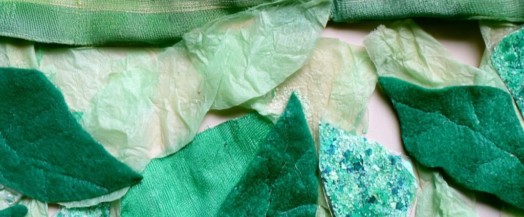
In elementary school, I spent an art class layering greens. Jungle green fabric with a subtle coordinating stripe, delicate spring green tissue paper so translucent you could see through it, pieces of chunky emerald felt cut into the shape of leaves, sparkling seafoam and teal glitter, all of it pressed and glued onto a piece of tagboard in what my teacher called a monochromatic collage. All shades of one color. Mono: one. Chroma: color. I didn’t think about the word at the time because I was too busy glittering.
That same week, I took a science quiz on monocots and dicots and got the answer wrong. I couldn’t remember which plants had one seed leaf and which had two. When I got the test back and saw my error, it clicked. Of course a monocot has one seed leaf. I’d been using mono in art class while missing it on a science quiz down the hall. The root moved between rooms, but I hadn’t learned yet that it could.
That stuck with me. Although my formal education is in English and linguistics, my mind naturally sought out scientific connections. Greek and Latin roots were the place where those two interests didn’t have to compete. They had the precision of formulas, a combinatorial logic where twenty roots could unlock hundreds of words, and they worked on both sides of the hallway.
I write about science now, and the roots have remained my constant companion. A few weeks ago I was researching an organism called Rapaza viridis for a blog post on endosymbiosis. I’d never heard of it, but since viridis is Latin for green (the same root that gives us “verdant”), I already guessed this creature had something to do with photosynthesis. It does. R. viridis is a single-celled predator that steals chloroplasts from the algae it hunts and uses them to photosynthesize.
Learning more about the organism, I came across the term “transient chimerism.” Also new to me, but also immediately legible. The trans in “transient” is the same as in “transparent” and “transport”: ‘across,’ ‘through.’ Something passing through. Chimera: the Greek monster stitched together from a lion, a goat, and a serpent. Put together: a temporary state of being made from parts of more than one organism. The roots came through for me once again.
This keeps happening. I’ll hit a term I’ve never seen, and its pieces already feel familiar. I could spend this entire blog walking through terms and showing you their roots, but definitions only stay interesting for so long (and my family already compares me to the father in My Big Fat Greek Wedding). What I’ve become more interested in is something the roots do beyond defining. The roots that end up in a name tend to carry more than a definition. They carry an interpretation, an argument about the thing itself.
For years, I wondered about apoptosis. I knew apo meant ‘away from.’ I could see ptosis maybe shared something with “asymptote,” but I couldn’t figure out what cell death had to do with calculus. In 1972, John Kerr, an Australian pathologist, had been studying a form of cell death that looked nothing like anything he’d seen before. For most of pathology’s history, the only cell death anyone studied was the kind that showed up when something had gone wrong: a cell damaged by injury or infection or toxins, dying violently. That kind of death had a name: necrosis, from the Greek nekros (‘corpse’). The cell swells, ruptures, spills its contents, and triggers inflammation. Studying liver tissue, Kerr noticed a second kind of death happening quietly alongside it, in cells that hadn’t been damaged at all. The cell death he was watching was the opposite. The cell shrank, and its contents condensed. It broke apart into tidy packages that neighboring cells quietly absorbed. No mess. The body had planned this.
Continue reading “Time Capsules in the Language of Science”