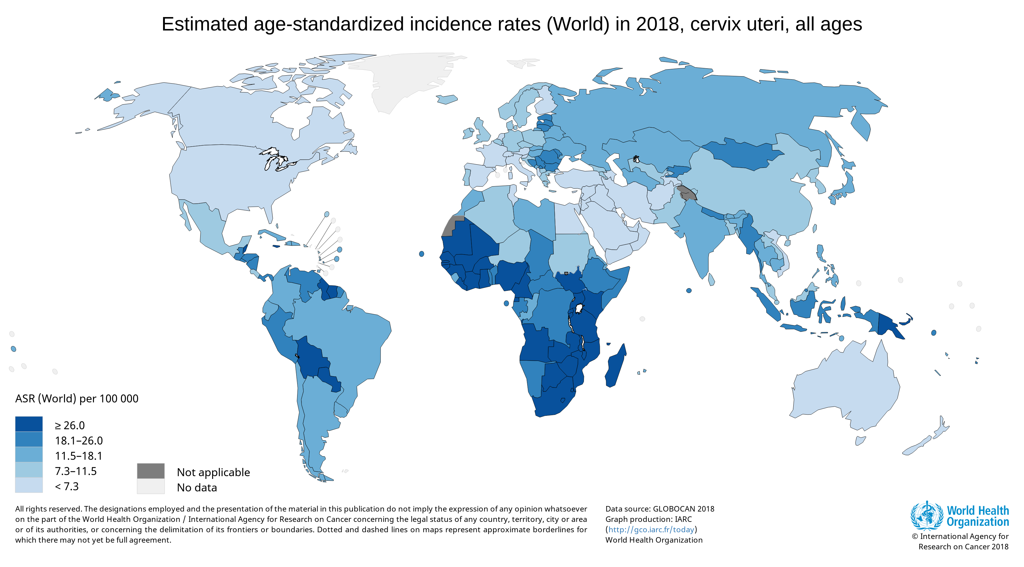
Cervical cancer is a major health problem for women, and it is currently the fourth most common cancer in women globally (1). A worldwide analysis of cancer estimates from the Global Cancer Observatory 2018 database showed that cervical cancer disproportionally affects lower-resource countries, on the basis of their Human Development Index; it was the leading cause of cancer-related death in women in many African countries (1).
Estimated cervical cancer global incidence rates from the GLOBOCAN 2018 database; image generated using IARC (http://go.iarc.fr/today).
Infection by human papillomavirus (HPV), a double-stranded DNA virus, is the leading cause of cervical cancer. Many types of HPV have been identified, and at least 14 high-risk HPV types are cancer-causing, according to a World Health Organization (WHO) fact sheet. Of these types, HPV-16 and HPV-18 are responsible for 70% of cervical cancers and pre-cancerous cervical lesions. HPV infection is sexually transmitted, most commonly by skin-to-skin genital contact. Although the majority of HPV infections are benign and resolve within a year or two, persistent infection in women, together with other risk factors, can lead to the development of cervical cancer [reviewed in (2)].
What animal can be found around the globe that outnumbers humans three to one? Gallus gallus domesticus, the humble chicken. The human appetite for eggs and lean meat drive demand for this feathered bird, resulting in a poultry population of over 20 billion.
Controversy over the origin of the domestic chicken (when, where and which species) have lead some researchers to look for that information in the genomes of contemporary chicken breeds and wild jungle fowl, the candidates from which chickens were derived. By sequencing over 600 genomes from Asian domestic poultry as well as 160 genomes from all four wild jungle fowl species and the five red jungle fowl subspecies, Wang et al. wanted to understand and identify the relationships and relatedness among these species and derive where domesticated chickens first arose.
In older people, low muscle mass is strongly associated with reduced functional capacity and an increased risk of disability. Myostatin is a negative regulator of muscle growth and has become an important target for pharmaceutical companies designing therapeutics to address age-associated muscle loss.
Anti-myostatin drugs increase muscle size and strength in preclinical studies. Fortetropin is a proteo-lipid complex made from fertilized egg yolk and shows anti-myostatin activity. When Fortetropin is provided as a supplement, lowered circulating myostatin levels are observed both in rodents and in young men. Fortetropin in combination with resistance exercise also lowers myostatin and increased lean body mass.
The development of NanoLuc® luciferase technology has provided researchers with new and better tools to study endogenous biology: how proteins behave in their native environments within cells and tissues. This small (~19kDa) luciferase enzyme, derived from the deep-sea shrimp Oplophorus gracilirostris, offers several advantages over firefly or Renilla luciferase. For an overview of NanoLuc® luciferase applications, see: NanoLuc® Luciferase Powers More than Reporter Assays.
The small size of NanoLuc® luciferase, as well the lack of a requirement for ATP to generate a bioluminescent signal, make it particularly attractive as a reporter for in vivo bioluminescent imaging, both in cells and live animals. Expression of a small reporter molecule as a fusion protein is less likely to interfere with the biological function of the target protein. NanoLuc® Binary Technology (NanoBiT®) takes this approach a step further by creating a complementation reporter system where one subunit is just 11 amino acids in length. This video explains how the high-affinity version of NanoBiT® complementation (HiBiT) works:
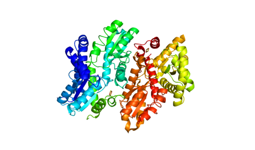
Human Ferrochelatase 2 angstrom crystal structure. Generated from 1HRK (RCSB PDB) using Pymol. Copyright: Sarah Wilson / CC BY-SA
Understanding how disease states arise from genetic variants is important for understanding disease resistance and progression. What can complicate our understanding of disease development is when two people have the same genetic variant, but only one has the disease. To investigate what might be happening with ferrochelatase (FECH) variant alleles that result in erythropoietic protoporphyria (EPP), scientists used next-generation sequencing (NGS) along with RNA analysis and DNA methylation testing to assess the FECH locus in 72 individuals from 24 unrelated families with EPP.
What is FECH and its relationship to EPP?
FECH is the gene for ferrochelatase, the last enzyme in the pathway that synthesizes heme. The inherited metabolic disorder, EPP, is caused when the activity of FECH is reduced to less than a third of normal levels thus, increasing the levels of protoporphyrin (PPIX) without metal in erythrocytes. The consequences of the low-metal PPIX include severe phototoxic skin reactions and hepatic injury due to PPIX accumulation in the liver.
How does FECH expression affect EPP?
The EPP disease state is not simply the lack of two functional FECH genes. Disease occurs with a hypomorphic allele, mutations in FECH that reduce its function, in trans to a null FECH allele. Researchers focused on three common variants called the GTC haplotype that are associated with expression quantitative trait loci (eQTL) that reduce FECH activity. Interestingly, these three variants have been found in trans, but researchers wanted to learn if there were individuals who were homozygous for the GTC allele and how EPP manifested for them.
Studying protein function in live cells is limited by the tools available to analyze the expression and interactions of those proteins. Although mass spectrometry and antibody-based protein detection are valuable technologies for protein analysis, both methods have drawbacks that limit the range of targets and contexts in which proteins can be investigated.
Mass spectrometry is often poor at detecting low-abundance proteins. Antibody-based techniques require high quality, specific antibodies, which can be difficult to impossible to acquire. Both methods require cell lysis, preventing real-time analysis and limiting the physiological relevance, and both methods can be limiting for higher-throughput analysis. While plasmid-based overexpression of tagged target proteins simplifies detection and can allow for real time analysis, protein levels don’t typically resemble endogenous levels. Overexpression also has the potential to create experimental artifacts or limit the dynamic range of an observed response.
In 2018, Promega R&D scientists published a paper in ACS Chemical Biology demonstrating the use of CRISPR/Cas9 to integrate the 11 amino acid, bioluminescent HiBiT tag directly into the genome to serve as an easily measured reporter for endogenous proteins. This provides a highly quantitative method for investigating cellular protein dynamics that sidesteps the need for cloning and other drawbacks to conventional methods, including the ability to measure changing protein dynamics in real-time. (For more details about CRISPR/Cas9 knock-in tagging and other applications, read this blog.)
While their findings showed that this method provides efficient and specific tagging of endogenous proteins, the research was limited to just five different proteins within a single signaling pathway in two cell lines. This left unanswered questions about whether this approach was scalable, had broader applications and how accurately the natural biology of the cells was represented.
RNA polymerase unwinds DNA strands for transcription.
Transcription is the production of RNA from a DNA sequence. It’s a necessary life process in most cells. Transcription performed in vitro is also a valuable technique for research applications—from gene expression studies to the development of RNA virus vaccines.
During transcription, the DNA sequence is read by RNA polymerase to produce a complimentary, antiparallel RNA strand. This RNA strand is called a primary transcript, often referred to as an RNA transcript. In vitro transcription is a convenient method for generating RNA in a controlled environment outside of a cell.
In vitro transcription offers flexibility when choosing a DNA template, with a few requirements. The template must be purified, linear, and include a double stranded promoter region. Acceptable template types are plasmids or cloning vectors, PCR products, synthetic oligos (oligonucleotides), and cDNA (complimentary DNA).
In vitro transcription is used for production of large amounts of RNA transcripts for use in many applications including gene expression studies, RNA interference studies (RNAi), generation of guide RNA (gRNA) for use in CRISPR, creation of RNA standards for quantification of results in reverse-transcription quantitative PCR (RT-qPCR), studies of RNA structure and function, labeling of RNA probes for blotting and hybridization or for RNA:protein interaction studies, and preparation of specific cDNA libraries, just to name a few!
In vitro transcription can also be applied in general virology to study the effects of an RNA virus on a cell or an organism, and in development and production of RNA therapeutics and RNA virus vaccines. The large quantity of viral RNA produced through in vitro transcription can be used as inoculation material for viral infection studies. Viral mRNA transcripts, typically coding for a disease-specific antigen, can be quickly created through in vitro transcription, and used in the production of vaccines and therapeutics.
Transcriptional activation of genes within the nucleus of eukaryotic cells occurs by a variety of mechanisms. Typically, these mechanisms rely on the interaction of regulatory proteins (transcriptional activators or repressors) with specific DNA sequences that control gene expression. Upon DNA binding, regulatory proteins also interact with other proteins that are part of the RNA polymerase II transcriptional complex.
One type of transcriptional activation relies on inducing a conformational change in chromatin, the DNA-protein complex that makes up each chromosome within a cell. In a broad sense, “extended” or loosely wound chromatin is more accessible to transcription factors and can signify an actively transcribed gene. In contrast, “condensed” chromatin hinders access to transcription factors and is characteristic of a transcriptionally inactive state. Acetylation of lysine residues in histones—the primary constituents of the chromatin backbone—results in opening up the chromatin and consequent gene activation. Disruption of histone acetylation pathways is implicated in many types of cancer (1).
Monitoring the use of performance-enhancing substances among athletes is complex and the requirements for tests and assays that detect use of such substances have changed significantly over the last few decades.
The haematological (blood) module of Athlete Biological Passport was adopted December 1, 2009 (ABP) by the World Anti-Doping Agency. The module sets out standard protocols to monitor doping of professional athletes by looking at changes in biological parameters, without relying on the detection of illegal compounds in body fluids. Such biological methods eliminate the need to develop and validate a test to detect every new compound that can be used for doping. The current version of the ABP, adopted in 2014, also adds monitoring of certain steroid use indicators from urine samples.
Blood doping which aims at increasing red blood cells so that more oxygen can be transported to muscles to increase stamina or performance is particularly difficult to detect. There are typically three ways that it is accomplished: use of erythropoietin (EPO) or synthetic oxygen carriers and blood transfusions. While transfusions of large volumes of blood or use of EPO can be detected, microdosing EPO or transfusing smaller volumes of packed red blood cells is much harder to detect.
Nicolas Leuenberger and colleagues at the Swiss Laboratory for Doping Analysis have developed a method to detect blood doping. In addition to addressing the detection of blood doping, his laboratory is also concerned about easing the transport and storage requirements for samples and ensuring that sample collection does not adversely affect athlete performance.
Improving Collection and Storage of Blood Samples
Because sample collection and storage are so critical to accurate test results, any new assays developed to detect blood doping benefit from ease of collection and storage. The Leuenberger laboratory investigated the use of the TAP™ Push Button collection device, which is billed as a simple method for blood collection that is easy to use and eliminates the need for painful needle sticks or finger pricks that can affect athlete performance. After TAP collection, 20µl of blood from the device was placed on to filter paper and dried (dried blood samples; DBS), which are much easier to store and transport from collection site to laboratory.
An RNA Biomarker for Blood Doping
Blood withdrawal and autologous transfusion or recombinant human EPO injection stimulate erythropoiesis and immature red blood cells can be distinguished based on their gene expression profiles. One of the genes that is expressed by immature red blood cells is aminoleuvulinate synthase 2, a gene that encodes an enzyme ALAS2 involved in the synthesis of heme, a pathway active during RBC maturation. RNA transcripts are unstable and tend to degrade rapidly, so isolating linear RNA transcripts from a collected sample can be difficult. However circular RNAs (circRNAs) are a class of RNA molecule produced by the backsplicing of pre-mRNAs that are high in abundance, quite stable and maintain cell-type specific expression. The Leuenberger laboratory developed a method for measuring the linear and circular forms of ALAS2 RNA in DBS to monitor erythropoiesis.
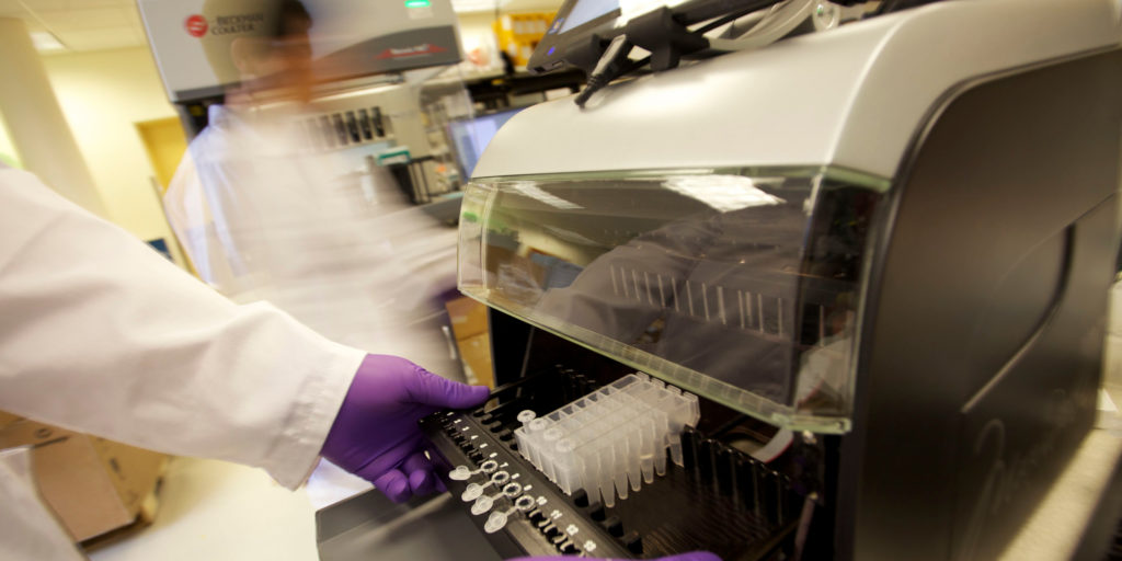
One of the greatest challenges in developing this protocol was achieving efficient RNA extraction from only 20ul of dried blood. Leuenberger and his colleagues adopted a two-step purification; beginning with a phenol:chloroform extraction on the DBS followed by a further purification on the Maxwell® RSC automated instrument, using the Maxwell RSC miRNA Serum and Plasma kit. Switching from a manual to an automated method for the second step was crucial. It reduced chances of contamination as well reduced pipetting errors, without compromising good quality and yield of RNA therefore contributing to assay reproducibility. To normalize volumes within the blood spot, the protocol uses RNA produced by housekeeping genes. The work to automate the assay has been published in Bioanalysis.
What’s Next
This protocol is being tested to see if microdosing of EPO or small transfusions can also be detected by monitoring ALAS2 RNA expression in DBS. The Swiss laboratory of Doping Analysis is also in the process of developing a method to detect gene doping by isolating plasmid DNA from whole blood samples, using the Maxwell® RSC.
Additionally, the collection and storage methods used have implications for the clinic, especially for patients that need routine blood monitoring. The ability to isolate circular RNAs shows promise in forensic applications to identify body fluids.
Want to know more about how the Maxwell® RSC can give you the freedom to focus on the work that interests you the most? To learn more, click here.
When I encounter my cat fixated on specific locations in my kitchen, her behavior shows me that she has heard some mice in those areas. In fact, mice have been attributed as a reason that cats became companions to humans. Mice start gathering and reproducing so cats followed the food source and hunted the rodents, thus endearing themselves to humans, who were storing food for their own use. However, new evidence described in Scientific Reports has shown that mice have been associated with humans even before grain storage was widespread. In fact, by making our dwellings comfortable, we also created an inviting place for mice to live.
XWe use cookies and similar technologies to make our website work, run analytics, improve our website, and show you personalized content and advertising. Some of these cookies are essential for our website to work. For others, we won’t set them unless you accept them. To learn more about our approach to Privacy we invite you to Read More
By clicking “Accept All”, you consent to the use of ALL the cookies. However you may visit Cookie Settings to provide a controlled consent.
We use cookies and similar technologies to make our website work, run analytics, improve our website, and show you personalized content and advertising. Some of these cookies are essential for our website to work. For others, we won’t set them unless you accept them. To find out more about cookies and how to manage cookies, read our Cookie Policy.
If you are located in the EEA, the United Kingdom, or Switzerland, you can change your settings at any time by clicking Manage Cookie Consent in the footer of our website.
Necessary cookies are absolutely essential for the website to function properly. These cookies ensure basic functionalities and security features of the website, anonymously.
Cookie
Duration
Description
cookielawinfo-checbox-analytics
11 months
This cookie is set by GDPR Cookie Consent plugin. The cookie is used to store the user consent for the cookies in the category "Analytics".
cookielawinfo-checbox-functional
11 months
The cookie is set by GDPR cookie consent to record the user consent for the cookies in the category "Functional".
cookielawinfo-checbox-others
11 months
This cookie is set by GDPR Cookie Consent plugin. The cookie is used to store the user consent for the cookies in the category "Other.
cookielawinfo-checkbox-advertisement
1 year
The cookie is set by GDPR cookie consent to record the user consent for the cookies in the category "Advertisement".
cookielawinfo-checkbox-necessary
11 months
This cookie is set by GDPR Cookie Consent plugin. The cookies is used to store the user consent for the cookies in the category "Necessary".
cookielawinfo-checkbox-performance
11 months
This cookie is set by GDPR Cookie Consent plugin. The cookie is used to store the user consent for the cookies in the category "Performance".
gdpr_status
6 months 2 days
This cookie is set by the provider Media.net. This cookie is used to check the status whether the user has accepted the cookie consent box. It also helps in not showing the cookie consent box upon re-entry to the website.
lang
This cookie is used to store the language preferences of a user to serve up content in that stored language the next time user visit the website.
viewed_cookie_policy
11 months
The cookie is set by the GDPR Cookie Consent plugin and is used to store whether or not user has consented to the use of cookies. It does not store any personal data.
Analytical cookies are used to understand how visitors interact with the website. These cookies help provide information on metrics the number of visitors, bounce rate, traffic source, etc.
Cookie
Duration
Description
SC_ANALYTICS_GLOBAL_COOKIE
10 years
This cookie is associated with Sitecore content and personalization. This cookie is used to identify the repeat visit from a single user. Sitecore will send a persistent session cookie to the web client.
vuid
2 years
This domain of this cookie is owned by Vimeo. This cookie is used by vimeo to collect tracking information. It sets a unique ID to embed videos to the website.
WMF-Last-Access
1 month 18 hours 24 minutes
This cookie is used to calculate unique devices accessing the website.
_ga
2 years
This cookie is installed by Google Analytics. The cookie is used to calculate visitor, session, campaign data and keep track of site usage for the site's analytics report. The cookies store information anonymously and assign a randomly generated number to identify unique visitors.
_gid
1 day
This cookie is installed by Google Analytics. The cookie is used to store information of how visitors use a website and helps in creating an analytics report of how the website is doing. The data collected including the number visitors, the source where they have come from, and the pages visted in an anonymous form.
Advertisement cookies are used to provide visitors with relevant ads and marketing campaigns. These cookies track visitors across websites and collect information to provide customized ads.
Cookie
Duration
Description
IDE
1 year 24 days
Used by Google DoubleClick and stores information about how the user uses the website and any other advertisement before visiting the website. This is used to present users with ads that are relevant to them according to the user profile.
test_cookie
15 minutes
This cookie is set by doubleclick.net. The purpose of the cookie is to determine if the user's browser supports cookies.
VISITOR_INFO1_LIVE
5 months 27 days
This cookie is set by Youtube. Used to track the information of the embedded YouTube videos on a website.
Performance cookies are used to understand and analyze the key performance indexes of the website which helps in delivering a better user experience for the visitors.
Cookie
Duration
Description
YSC
session
This cookies is set by Youtube and is used to track the views of embedded videos.
_gat_UA-62336821-1
1 minute
This is a pattern type cookie set by Google Analytics, where the pattern element on the name contains the unique identity number of the account or website it relates to. It appears to be a variation of the _gat cookie which is used to limit the amount of data recorded by Google on high traffic volume websites.