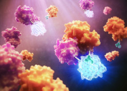
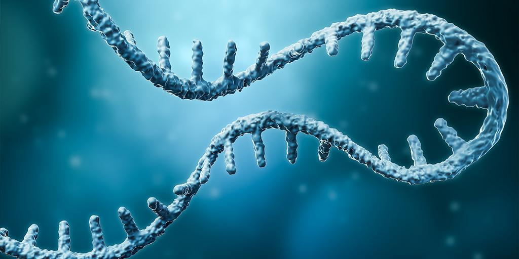
Almost 90% of the human genome is transcribed into RNA, but only 3% is ultimately translated into a protein. Some non-translated RNA is thought to be useless, while some play a significant yet often mysterious role in cancer and other diseases. Despite its abundance and biological significance, RNA is rarely the target of therapeutics.
“We say it’s undruggable, but I would say that ‘not-yet-drugged’ is a better way to put it,” says Amanda Garner, Associate Professor of Medicinal Chemistry at the University of Michigan. “We know that RNA biology is important, but we don’t yet know how to target it.”
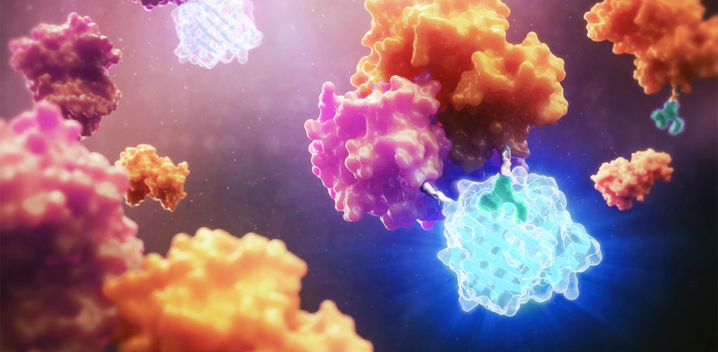
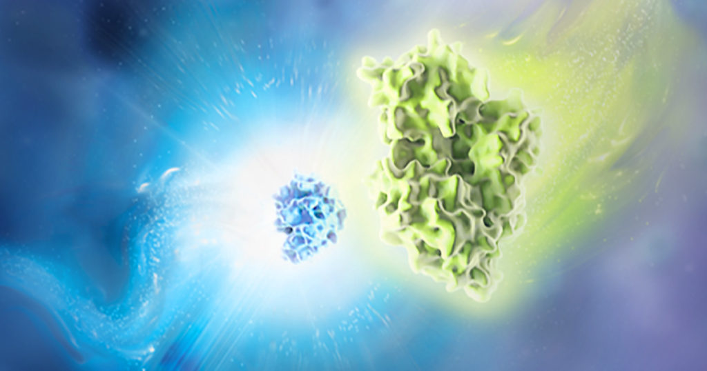
Amanda’s lab develops systems to study RNA biology. She employs a variety of approaches to analyze the functions of different RNAs and study their interactions with proteins. Her lab recently published a paper describing a novel method for studying RNA-protein interactions (RPI) in live cells. Amanda says that with the right tools, RPI could become a critical target for drug discovery.
“It’s amazing that current drugs ever work, because they’re all based on really old approaches,” Amanda says. “This isn’t going to be like developing a small molecule kinase inhibitor. It’s a whole new world.”
Continue reading “RNA-Protein Interactions: A New Frontier for Drug Discovery”