Author: Sara Klink
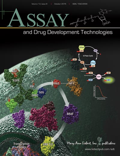
A Better GTPase Assay for Drug Development
The path to drug development is strewn with obstacles: Identifying targets; configuring assays to help identify targets or drugs; uncovering the right compound to affect the selected target without off-target effects and screening multiple compounds to eliminate or identify potential drugs. Without the right tools, compounds or target, identifying potential disease therapies becomes nearly impossible.
When it comes to a drug target for cancer, the Ras protein family is at the top of the list because the proteins are expressed ubiquitiously and found mutated in many types of cancer. Because Ras proteins are involved in transducing signals from the surface of cells, many of the resulting mutations produce an activated Ras, inducing uncontrolled expression of the genes that Ras controls. Ras proteins are small GTPases (20–25kDa) that comprise a larger superfamily of proteins divided into five subfamilies: Ras, Rho, Rab, Arf, and Ran. These proteins control diverse cellular activities, including cellular differentiation, proliferation, cell division, nuclear import and export, and vesicle transport. GTPases are guanosine-nucleotide-binding proteins with affinity for GDP or GTP and are able to hydrolyze GTP. When bound to GTP, GTPases are active (turned on) and interact with downstream proteins in the signaling cascade. When GTPases are bound to GDP, the proteins are inactivated (turned off) and no longer transduce signals.
Continue reading “A Better GTPase Assay for Drug Development”Yersinia pestis Reveals More Secrets From the Grave

Fridays are generally reserved for fun posts to share prior to the weekend. As we all know, fun is relative and to me, the latest news about how long Yersinia pestis has been entwined with human history is intriguing. I enjoy writing about the latest historical finding of Y. pestis even if I do earn a black reputation among my blogging colleagues (pun intended). Therefore, as soon as I saw the Cell article about Y. pestis found in Bronze age human teeth, I knew my blog topic was at hand.
Y. pestis has long been suspected in several plagues that occurred in the last two millennia. Publications in 2011 and 2013 used DNA extracted from teeth of human remains dated to the 14th century Black Death and 6th century Plague of Justinian to confirm Y. pestis was the causative agent in those devastating plagues. These results beg the question: How long has Y. pestis been infecting humans? The phylogenic trees generated from recent studies suggested Y. pestis has been with humans for as little as 2,600 years and as long as and 28,000 years. Equipped with these DNA-based tools, Rasmussen et al. asked if they could find evidence of Y. pestis in older human remains.
Continue reading “Yersinia pestis Reveals More Secrets From the Grave”A Potential Single-Tube Multiplex Assay for Detecting Dengue Virus in the Field
In areas of the world where the electricity is intermittent, resources are limited and transporting bulky equipment and reagents that are sensitive to temperature fluctuations is difficult, diagnosis of viruses like dengue can be challenging. If you could reduce or eliminate the need for electricity dependent equipment for diagnostic assays without sacrificing sensitivity or specificity, it would be a boon to field workers. An article published in PLOS ONE describes how researchers developed a multiplex isothermal amplification method that could assess a potential dengue infection with a visual real-time or endpoint detection in a single tube.

Friday Cartoon Fun: Is It Drawn or Is It Real?
I always enjoy Ed Himelblau’s cartoons, but one that makes me chuckle every time I see it is the following:

I am sure our readers that enjoy coffee can empathize.
Recently, our Swiss branch had fun with a number of the cartoons from our Cartoon Lab archive and recreated the cartoon in real life:
What do you think?
All You Need is Pla (for Pneumonic Plague)

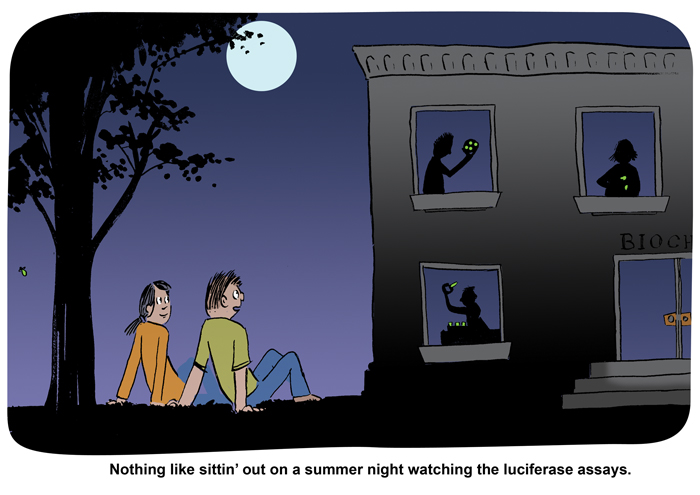
Friday Cartoon Post: Do You See the Assays Glowing?
As a tribute to the fireflies lighting up the night during July evenings in Wisconsin and the reporter gene assays they inspire, I wanted to share a special Ed Himelblau cartoon:
Developing a Model System to Test Ketamine Toxicity
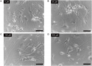
Cells were treated with 0μM (Panel A), 20μM (Panel B), 100μM (Panel C) or 500μM (Panel D) ketamine for 24 hours. Scale bar = 50μm. From Ito, H., Uchida, T. and Makita, K. (2015) Ketamine causes mitochondrial dysfunction in human induced pluripotent stem cell-derived neurons. PLOS ONE 10, e0128445.
doi:10.1371/journal.pone.0128445.g002
Cancer Detection on a Chip?
Cloning Tips for Restriction Enzyme-Digested Vectors and Inserts