You are studying the effects of a compound(s) on your cells. You want to know how the compound affects cell health over a period of hours, or even days. Real-time assays allow you to monitor cell viability, cytotoxicity and apoptosis continuously, to detect changes over time.
Why use a real-time assay? A real-time assay enables you to repeatedly measure specific events or conditions over time from the same sample or plate well. Repeated measurement is possible because the cells are not harmed by real-time assay reagents. Real-time assays allow you to collect data without lysing the cells.
Advantages of Real-Time Measurement Real-time assays allow you to:
When someone is admitted to a hospital for an illness, the hope is that medical care and treatment will help them them feel better. However, nosocomial infections—infections acquired in a health-care setting—are becoming more prevalent and are associated with an increased mortality rate worldwide. This is largely due to the misuse of antibiotics, allowing some bacteria to become resistant. Furthermore, when an antibiotic wipes out the “good” bacteria that comprise the human microbiome, it leaves a patient vulnerable to opportunistic infections that take advantage of disruptions to the gut microbiota.
One such bacteria, Clostridium difficile, is of growing concern world-wide since it is resistant to many different antibiotics. When a patient is treated with an antibiotic, C. difficile can thrive in the intestinal tract without other bacteria populating the gut. C. difficile infection is the leading cause of antibiotic-associated diarrhea. While symptoms can be mild, aggressive infection can lead to pseudomembranous colitis—a severe inflammation of the colon which can be life-threatening.
C. difficile causes disease by releasing two large toxins, TcdA and TcdB. Understanding the role these toxins play in colonic disease is important for treatment strategies. However, most published research data only report the effects of the toxins independently. A 2016 study demonstrated a method of comparing the toxins side-by-side using the same time points and cell assays to investigate the role each toxin plays in the cell death that leads to disease of the colon. Continue reading “A Tale of Two Toxins: the mechanisms of cell death in Clostridium difficile infections”
What if you could uncover a small but significant cellular response as your population of cells move toward apoptosis or necrosis? What if you could view the full picture of cellular changes rather than a single snapshot at one point? You can! There are real-time assays that can look at the kinetics of changes in cell viability, apoptosis, necrosis and cytotoxicity—all in a plate-based format. Seeking more information? Multiplex a real-time assay with endpoint analysis. From molecular profiling to complementary assays (e.g., an endpoint cell viability assay paired with a real-time apoptosis assay), you can discover more information hidden in the same cells during the same experiment.
Whether your research involves screening a panel of compounds or perturbing a regulatory pathway, a more complete picture of cellular changes gives you the benefit of more data points for better decision making. Rather than assessing the results of your experiment using a single time point, such as 48 hours, you could monitor cellular changes at regular intervals. For instance, a nonlytic live-cell reagent can be added to cultured cells and measurements taken repeatedly over time. Pairing a real-time cell health reagent with a detection instrument that can maintain the cells at the correct temperature means you can automate the measurements. These repeated measurements over time reveal the kinetic changes in the cells you are testing, giving a real-time status update of the cellular changes from the beginning to the end of your experiment. Continue reading “Reveal More Biology: How Real-Time Kinetic Cell Health Assays Prove Their Worth”
In today’s post, guest blogger, Martha O’Brien, PhD, provides a preview of her upcoming AAI poster and block symposium talk on the inflammasome, caspase-1 activity and pyroptosis.
Schematic of the Caspase-Glo 1 Inflammasome Assay.
Responding rapidly to microbial pathogens and damage-associated molecular markers is critical to our innate immune system. Caspase-1 is pivotal in this process leading to processing and release of essential cytokines and an immunogenic form of cell death, termed pyroptosis. Upon sensing pathogen-associated and damage-associated molecular patterns (PAMPs and DAMPs), innate immune cells form inflammasome protein complexes that recruit and activate caspase-1 (canonical inflammasomes). In addition, other inflammatory caspases, 4 and 5 in humans and 11 in mice, directly bind bacterial lipopolysaccharides (LPS), triggering pyroptosis (non-canonical inflammasome). LPS-triggered non-canonical inflammasomes in mice and humans ultimately lead to canonical inflammasome engagement and caspase-1 activation (1–3). Caspase-1 was originally termed interleukin converting enzyme (ICE) for its well-established role in processing IL-1ß and IL-18, two important inflammation cytokines. How caspase-1 mediates pyroptosis is less well understood, but is beginning to be delineated. Recently, a substrate of the inflammatory caspases, gasdermin D, was identified and its processed fragment, gasdermin-N domain, was shown to be required for pyroptosis in non-canonical inflammasome circumstances (4, 5). The precise role of gasdermin D in canonical inflammasome-triggered pyroptosis is still under investigation. Linking inflammatory caspases directly to pyroptosis is a notable step in understanding the mechanism of this important form of cell death.
Pyroptosis is clearly one means of releasing processed IL-1ß and IL-18 from the cell. However depending on the cell type and stimulus, there is evidence for inflammasome engagement, caspase-1 activation, and release of IL-1ß in the absence of cell death (6, 7). On the flip-side there is also evidence for caspase-1 mediated pyroptosis that helps clear bacteria, independent of IL-1ß and IL-18 involvement (8). To enable further studies on the inflammasome and in particular, assessing the connections between caspase-1 activation, pyroptosis, and cytokine release, Promega developed a new tool to conveniently monitor caspase-1 activation, the Caspase-Glo® 1 Inflammasome Assay. This bioluminescent, plate-based assay is used to measure caspase-1 activity directly in cell cultures or to monitor released caspase-1 activity in culture medium from treated cells. This flexibility allows easy multiplexing to monitor all three outcomes of inflammasome stimulation; caspase-1 activity, pyroptosis, and release of IL-1ß and IL-18. Caspase-1 activation typically is monitored indirectly with western blots of processed caspase-1. Now the activity of the enzyme can be monitored directly, providing accurate information on temporal aspects of the inflammasome. The assay can be readily combined with real-time measures of cell death (e.g., CellTox™ Green Cytotoxicity Assay) and some of the culture medium can be removed for IL-1ß/IL-18 assessment, leaving the cells and remaining culture medium for caspase-1 activity measurements.
Based on the Illuminations article by Dr. Terry Riss, from our Cellular Analysis group.
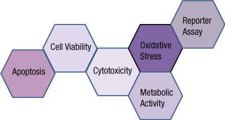
Choosing the most appropriate cell health assay for your experiment can be difficult. There are several factors to consider when choosing an assay: the question you are asking, the nature of your sample, the number of samples being tested, the required sensitivity, the nature of the sample, the plates and plate readers and the reagent costs.
What question are you asking?
The first, and perhaps most important factor to consider, is the question you need answered. What do you want to know at the end of the experiment? There are cell health assays available that specifically detect the number of living cells, the number of dead cells, and for assessing stress response mechanisms or pathways that may lead to cell death. Matching the assay endpoint to the information you need is vital to choosing the appropriate cell health assay. Continue reading “Choosing the Right Cell Health Assay”
You often need several pieces of information to really understand what is happening within a cell or population of cells. If your cells are not proliferating, are they dying? Or, are you seeing cytostasis? If they are dying, what is the mechanism? Is it apoptosis or necrosis? If you are seeing apoptosis, what is the pathway: intrinsic or extrinsic?
If you are measuring expression of a reporter gene and you see a decrease in expression, is that decrease due to transfection inefficiencies, cytotoxicity, or true down regulation of your reporter gene?
To investigate these multiple parameters, you can run assays in parallel, but that requires more sample, and sample isn’t always abundant.
Multiplexing assays allows you to obtain information about multiple parameters or events (e.g., reporter gene expression and cell viability; caspase-3 activity and cell viability) from a single sample. Multiplexing saves sample, saves time and gives you a more complete picture of the biology that is happening with your experimental sample.
What information do you need about your cells to complete the picture?
Multiplexing assay reagents to measure biomarkers in the same sample has often been considered an application only accomplished with antibodies or dyes and sophisticated detection instrumentation. However, Promega has developed microwell plate based assays for cells in culture that allow multiplexed detection of biomarkers in the same sample well using standard multimode multiwell plate readers. Continue reading “Piecing the Puzzle Together: Using Multiple Assays to Better Understand What Is Happening with Your Cells”
Life is complicated. So is death. And when the cells in your multiwell plate die after compound treatment, it’s not enough to know that they died. You need to know how they died: apoptosis or necrosis? Or, have you really just reduced viability, rather than induced death? Is the cytotoxicity you see dose-dependent? If you look earlier during drug treatment of your cells, do you see markers of apoptosis? If you wait longer, do you observe necrosis? If you reduce the dosage of your test compound, is it still cytotoxic? Continue reading “Describing Life and Death in the Cell”
XWe use cookies and similar technologies to make our website work, run analytics, improve our website, and show you personalized content and advertising. Some of these cookies are essential for our website to work. For others, we won’t set them unless you accept them. To learn more about our approach to Privacy we invite you to Read More
By clicking “Accept All”, you consent to the use of ALL the cookies. However you may visit Cookie Settings to provide a controlled consent.
We use cookies and similar technologies to make our website work, run analytics, improve our website, and show you personalized content and advertising. Some of these cookies are essential for our website to work. For others, we won’t set them unless you accept them. To find out more about cookies and how to manage cookies, read our Cookie Policy.
If you are located in the EEA, the United Kingdom, or Switzerland, you can change your settings at any time by clicking Manage Cookie Consent in the footer of our website.
Necessary cookies are absolutely essential for the website to function properly. These cookies ensure basic functionalities and security features of the website, anonymously.
Cookie
Duration
Description
cookielawinfo-checbox-analytics
11 months
This cookie is set by GDPR Cookie Consent plugin. The cookie is used to store the user consent for the cookies in the category "Analytics".
cookielawinfo-checbox-functional
11 months
The cookie is set by GDPR cookie consent to record the user consent for the cookies in the category "Functional".
cookielawinfo-checbox-others
11 months
This cookie is set by GDPR Cookie Consent plugin. The cookie is used to store the user consent for the cookies in the category "Other.
cookielawinfo-checkbox-advertisement
1 year
The cookie is set by GDPR cookie consent to record the user consent for the cookies in the category "Advertisement".
cookielawinfo-checkbox-necessary
11 months
This cookie is set by GDPR Cookie Consent plugin. The cookies is used to store the user consent for the cookies in the category "Necessary".
cookielawinfo-checkbox-performance
11 months
This cookie is set by GDPR Cookie Consent plugin. The cookie is used to store the user consent for the cookies in the category "Performance".
gdpr_status
6 months 2 days
This cookie is set by the provider Media.net. This cookie is used to check the status whether the user has accepted the cookie consent box. It also helps in not showing the cookie consent box upon re-entry to the website.
lang
This cookie is used to store the language preferences of a user to serve up content in that stored language the next time user visit the website.
viewed_cookie_policy
11 months
The cookie is set by the GDPR Cookie Consent plugin and is used to store whether or not user has consented to the use of cookies. It does not store any personal data.
Analytical cookies are used to understand how visitors interact with the website. These cookies help provide information on metrics the number of visitors, bounce rate, traffic source, etc.
Cookie
Duration
Description
SC_ANALYTICS_GLOBAL_COOKIE
10 years
This cookie is associated with Sitecore content and personalization. This cookie is used to identify the repeat visit from a single user. Sitecore will send a persistent session cookie to the web client.
vuid
2 years
This domain of this cookie is owned by Vimeo. This cookie is used by vimeo to collect tracking information. It sets a unique ID to embed videos to the website.
WMF-Last-Access
1 month 18 hours 24 minutes
This cookie is used to calculate unique devices accessing the website.
_ga
2 years
This cookie is installed by Google Analytics. The cookie is used to calculate visitor, session, campaign data and keep track of site usage for the site's analytics report. The cookies store information anonymously and assign a randomly generated number to identify unique visitors.
_gid
1 day
This cookie is installed by Google Analytics. The cookie is used to store information of how visitors use a website and helps in creating an analytics report of how the website is doing. The data collected including the number visitors, the source where they have come from, and the pages visted in an anonymous form.
Advertisement cookies are used to provide visitors with relevant ads and marketing campaigns. These cookies track visitors across websites and collect information to provide customized ads.
Cookie
Duration
Description
IDE
1 year 24 days
Used by Google DoubleClick and stores information about how the user uses the website and any other advertisement before visiting the website. This is used to present users with ads that are relevant to them according to the user profile.
test_cookie
15 minutes
This cookie is set by doubleclick.net. The purpose of the cookie is to determine if the user's browser supports cookies.
VISITOR_INFO1_LIVE
5 months 27 days
This cookie is set by Youtube. Used to track the information of the embedded YouTube videos on a website.
Performance cookies are used to understand and analyze the key performance indexes of the website which helps in delivering a better user experience for the visitors.
Cookie
Duration
Description
YSC
session
This cookies is set by Youtube and is used to track the views of embedded videos.
_gat_UA-62336821-1
1 minute
This is a pattern type cookie set by Google Analytics, where the pattern element on the name contains the unique identity number of the account or website it relates to. It appears to be a variation of the _gat cookie which is used to limit the amount of data recorded by Google on high traffic volume websites.
 When someone is admitted to a hospital for an illness, the hope is that medical care and treatment will help them them feel better. However, nosocomial infections—infections acquired in a health-care setting—are becoming more prevalent and are associated with an increased mortality rate worldwide. This is largely due to the misuse of antibiotics, allowing some bacteria to become resistant. Furthermore, when an antibiotic wipes out the “good” bacteria that comprise the human microbiome, it leaves a patient vulnerable to opportunistic infections that take advantage of disruptions to the gut microbiota.
 What if you could uncover a small but significant cellular response as your population of cells move toward apoptosis or necrosis? What if you could view the full picture of cellular changes rather than a single snapshot at one point? You can! There are real-time assays that can look at the kinetics of changes in cell viability, apoptosis, necrosis and cytotoxicity—all in a plate-based format. Seeking more information? Multiplex a real-time assay with endpoint analysis. From molecular profiling to complementary assays (e.g., an endpoint cell viability assay paired with a real-time apoptosis assay), you can discover more information hidden in the same cells during the same experiment.
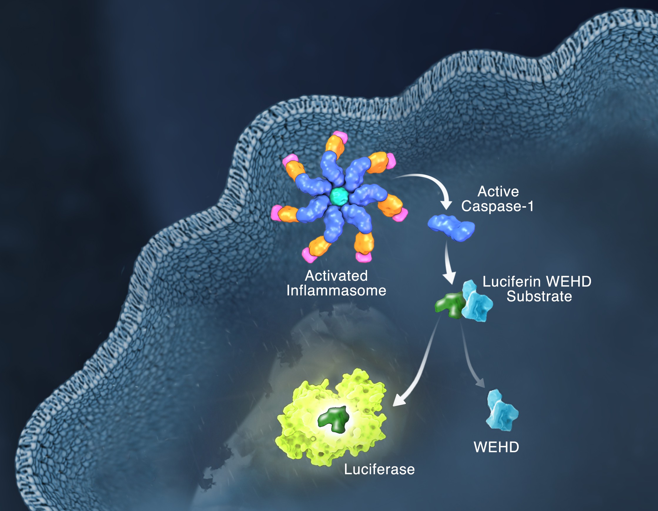
 Life is complicated. So is death. And when the cells in your multiwell plate die after compound treatment, it’s not enough to know that they died. You need to know how they died: apoptosis or necrosis? Or, have you really just reduced viability, rather than induced death? Is the cytotoxicity you see dose-dependent? If you look earlier during drug treatment of your cells, do you see markers of apoptosis? If you wait longer, do you observe necrosis? If you reduce the dosage of your test compound, is it still cytotoxic?