Detecting Disulfide Bond Shuffling in Biologics Using Trypsin Platinum

Biologic therapeutics such as monoclonal antibodies and biosimilars are complex proteins that are susceptible to post-translational modifications (PTMs). These chemical modifications can affect the performance and activity of the biologic, potentially resulting in decreased potency and increased immunogenicity. Such modifications include glycosylation, deamidation, oxidation and disulfide bond shuffling. These PTMs can be signs of protein degradation, manufacturing issues or improper storage. Several of these modifications are well characterized, and methods exist for detecting them during biologic manufacture. However, disulfide shuffling is not particularly well characterized for biologics, and no methods exist to easily detect and quantify disulfide bond shuffling in biologics.

Disulfide bond shuffling occurs when the S-S linkage is not between a Cys and its normal partner
Disulfide bonds are important for protein conformation and function

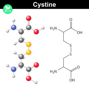
Normally the cysteines in a protein will pair with a predictable or “normal” partner residue either within a polypeptide chain or between two polypeptide chains when they form disulfide bonds. These normal disulfide bonds are important for final protein conformation and stability. Indeed, disulfide bonds are considered an important quality indicator for biologics.

In a recently published study, Coghlan and colleagues designed a semi-automated method for characterizing disulfide bond shuffling on two IgG1 biologics: rituximab (originator drug Rituxan® and biosimilar Acellbia®) and bevacizumab (originator Avastin® and biosimilar Avegra®).

Continue reading “Detecting Disulfide Bond Shuffling in Biologics Using Trypsin Platinum”

Why Wait? Sample Prep/Protein Digestion in as Little as 30 Minutes!

While many proteases are used in bottom-up mass spectrometric (MS) analysis, trypsin (4,5) is the de facto protease of choice for most applications. There are several reasons for this: Trypsin is highly efficient, active, and specific. Tryptic peptides produced after proteolysis are ideally suited, in terms of both size (350–1,600 Daltons) and charge (+2 to +4), for MS analysis. One significant drawback to trypsin digestion is the long sample preparation times, which typically range from 4 hours to overnight for most protocols. Achieving efficient digestion usually requires that protein substrates first be unfolded either with surfactants or denaturants such as urea or guanidine. These chemical additives can have negative effects, including protein modification, inhibition of trypsin or incompatibility with downstream LC-MS/MS. Accordingly, additional steps are typically required to remove these compounds prior to analysis.

Continue reading “Why Wait? Sample Prep/Protein Digestion in as Little as 30 Minutes!”

Optimization of Alternative Proteases for Bottom-Up Proteomics

Alternate Proteases Cover

Bottom-up proteomics focuses on the analysis of protein mixtures after enzymatic digestion of the proteins into peptides. The resulting complex mixture of peptides is analyzed by reverse-phase liquid chromatography (RP-LC) coupled to tandem mass spectrometry (MS/MS). Identification of peptides and subsequently proteins is completed by matching peptide fragment ion spectra to theoretical spectra generated from protein databases.

Trypsin has become the gold standard for protein digestion to peptides for shotgun proteomics. Trypsin is a serine protease. It cleaves proteins into peptides with an average size of 700-1500 daltons, which is in the ideal range for MS (1). It is highly specific, cutting at the carboxyl side of arginine and lysine residues. The C-terminal arginine and lysine peptides are charged, making them detectable by MS. Trypsin is highly active and tolerant of many additives.

Even with these technical features, the use of trypsin in bottom-up proteomics may impose certain limits in the ability to grasp the full proteome, Tightly-folded proteins can resist trypsin digestion. Post-translational modifications (PTMs) present a different challenge for trypsin because glycans often limit trypsin access to cleavage sites, and acetylation makes lysine and arginine residues resistant to trypsin digestion.

To overcome these problems, the proteomics community has begun to explore alternative proteases to complement trypsin. However, protocols, as well as expected results generated when using these alternative proteases have not been systematically documented.

In a recent reference (2), optimized protocols for six alternative proteases that have already shown promise in their applicability in proteomics, namely chymotrypsin, Lys-C, Lys-N, Asp-N, Glu-C and Arg-C have been created.

Data describe the appropriate MS data analysis methods and the anticipated results in the case of the analysis of a single protein (BSA) and a more complex cellular lysate (Escherichia coli). The digestion protocol presented here is convenient and robust and can be completed in approximately in 2 days.


Try a sample of high-efficiency Trypsin Platinum today!

Visit our website for more on Trypsin Platinum, Mass Spectrometry Grade, with enhanced proteolytic efficiency and superior autoproteolytic resistance.


Reference

  1. Laskay, U. et al. (2013) Proteome Digestion Specificity Analysis for the Rational Design of Extended Bottom-up and middle-down proteomics experiments. J of Proteome Res. 12, 5558–69.
  2. Giansanti, P. et. al. (2016) Six alternative protease for mass spectrometry based proteomics beyond trypsin. Nat. Protocols 11, 993–6

Related Posts