Designing BET(ter) Inhibitors to Guide Therapy for Cancer and Inflammatory Diseases

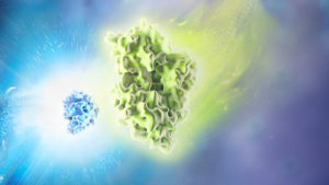
bet proteins brd nanoluc

Transcriptional activation of genes within the nucleus of eukaryotic cells occurs by a variety of mechanisms. Typically, these mechanisms rely on the interaction of regulatory proteins (transcriptional activators or repressors) with specific DNA sequences that control gene expression. Upon DNA binding, regulatory proteins also interact with other proteins that are part of the RNA polymerase II transcriptional complex.

One type of transcriptional activation relies on inducing a conformational change in chromatin, the DNA-protein complex that makes up each chromosome within a cell. In a broad sense, “extended” or loosely wound chromatin is more accessible to transcription factors and can signify an actively transcribed gene. In contrast, “condensed” chromatin hinders access to transcription factors and is characteristic of a transcriptionally inactive state. Acetylation of lysine residues in histones—the primary constituents of the chromatin backbone—results in opening up the chromatin and consequent gene activation. Disruption of histone acetylation pathways is implicated in many types of cancer (1).

Continue reading “Designing BET(ter) Inhibitors to Guide Therapy for Cancer and Inflammatory Diseases”

Protein:DNA Interactions—High-Throughput Analysis

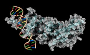
Copyright: molekuul / 123RF Stock Photo

Protein-DNA interactions are fundamental processes in gene regulation in a living cells. These interactions affect a wide variety of cellular processes including DNA replication, repair, and recombination. In vivo methods such as chromatin immunoprecipitation (1) and in vitro electrophoretic mobility shift assays (2) have been used for several years in the characterization of protein-DNA interactions. However, these methods lack the throughput required for answering genome-wide questions and do not measure absolute binding affinities. To address these issues a recent publication (3) presented a high-throughput micro fluidic platform for Quantitative Protein Interaction with DNA (QPID). QPID is an microfluidic-based assay that cam perform up to 4096 parallel measurements on a single device.

The basic elements of each experiment includes oligonucleotides that were synthesized and hybridized to a Cy5-labeled primer and extended using Klenow. All transcription factors that were evaluated contained a 3’HIS and 5’ cMyc tag and were expressed in rabbit reticulocyte coupled transcription and translation reaction (TNT® Promega). Expressed proteins are loaded onto to the QIPD device and immobilized. In the DNA binding assay the fluorescent DNA oligonucleotides are incubated with the immobilized transcription factors and fluorescent images taken. To validate this concept the binding of four different transcription factor complexes to 32 oligonucleotides at 32 different concentrations was characterized in a single experiment. In a second application, the binding of ATF1 and ATF3 to 128 different DNA sequences at different concentrations were analyzed on a single device.

Literature Cited

  1. Ren, B. et al. (2007) Genome-wide mapping of in vivo protein-DNA binding proteins. Science 316, 1497–502.
  2. Garner, M.M. (1981) A gel electrophoresis method for quantifying the binding of proteins to specific DNA regions. Nuc. Acids. Res. 9, 3047-60.
  3. Glick,Y et al. (2016) Integrated microfluidic approach for quantitative high throughput measurements of transcription factor binding affinities. Nuc. Acid Res. 44, e51.

Decorating Histones and Their Tails

There are at least two tail stories associated with big scientific discoveries. One is Darwin’s story about the tail loss during human evolution process. The other story is associated with discovery of benzene ring structure. In his creative dream Kekule saw the snake (the linear carbon chain) eating its own tail. Even better representation of benzene structure is the comic image of six monkeys holding each other hands and tails. Nowadays, the most popular scientific story in the field of epigenetics is the story of histones and their tails. This time instead of monkey or snake, an elephant is the animal whose characteristics allegorically represent epigenetics.

elephants_web

We all know how the histone octamer wrapped with DNA represents a nucleosome – the first unit of chromatin formation. Histones, which are basic due to numerous arginines and lysines, easily attract negatively charged DNA and in that way facilitate formation of nucleosome. The nature of two materials is important but not sufficient for such complex biological function like efficient packaging of DNA and regulation of gene expression. For that reason both the DNA and histones are decorated by numerous chemical groups.

Post-translational modifications (PTMs) of histones and histone variants themselves can cause alternation of net charge, changes histone dynamics and interaction with other chromatin proteins. The extreme complexity of interactions that can be achieved by histone modifications inspired Jenuwein and Allis to launch an idea of “histone or epigenetic code”. Core histones consist of a N- terminal tail, the globular portion and a C terminus. PMTs were discovered first on the N-terminal tail of core histones. However, the logical question was: Are only the tails decorated or are there more?” Continue reading “Decorating Histones and Their Tails”