Updated 4/29/2022 by AnnaKay Kruger
Viruses are both fascinating and terrifying. Stealthy, insidious and often deadly, they turn our own cells against us. Over the past year, we have all had a firsthand view of what a new and unknown virus can do. The SARS-CoV-2 virus has caused a global pandemic, and left scientists and medical professionals scrambling to unravel its mysteries and find ways to stop it.
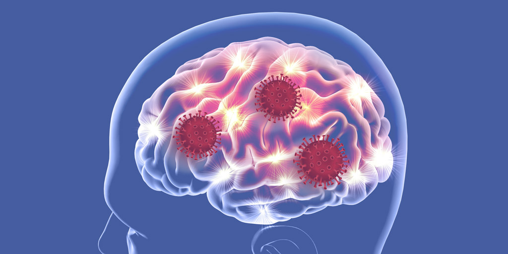
COVID-19 is considered a respiratory disease, but we know that the SARS-CoV-2 virus can affect other systems in the body including the vascular and central nervous systems. In fact, some of the most noted symptoms of SARS-CoV-2 infection, headache, and the loss of the sense of taste and smell, are neurological— not respiratory— symptoms.
Recently, researchers publishing in the Journal of Experimental Medicine (1) described their work to help better understand the ability of SARS-CoV-2 to infect the brain. First, they used human brain organoids as an in vitro method to evaluate if the virus could infect neurons and leverage their cellular processes to replicate. Second, they used mice that were genetically engineered to over express human ACE2—the protein SARS-CoV-2 binds to enter lung cells— to investigate in vivo infection of neurological tissue. Finally, they looked for SARS-CoV-2 in the brains of three individuals who had died from COVID-19.
SARS-CoV-2 Can Infect Neuronal Tissue in vitro using ACE2
Using human brain organoids, the researchers demonstrated SARS-CoV-2 infection of neurons and showed that the virus was able to use these cells to replicate. Upon infection, it appears that the virus boosts the metabolism of the infected cells and may cause localized hypoxic regions that result in the death of uninfected neighbor cells. Although mRNA levels of ACE2 appear to be very low in the central nervous system, the researchers found widespread ACE2 protein expression in the brain organoids using immunofluorescent staining. Additionally, postmortem human brains were stained for neurons and ACE2, and the ACE2 staining colocalized to neurons in the cortical grey matter. They further showed the requirement by blocking ACE2 using antibodies and cerebrospinal fluid containing anti-viral antibodies. When the ACE2 protein was blocked, the virus was prevented from infecting the brain organoids. These findings suggest that, just like in the lungs, SARS-CoV-2 requires ACE2 to successfully infect neuronal cells. It also appears that ACE2 is expressed in vitro and in vivo in the central nervous system.
SARS-CoV-2 Can Infect Neuronal Tissue Expressing Human ACE2 in vivo
The researchers tested the neural invasive capabilities of SARS-CoV-2 in an in vivo system using mice that were genetically engineered to over express human ACE2. They used labeled antibodies to track the distribution of the virus following intranasal infection and found that the neural cells of the forebrain were widely infected, and the cortex was unevenly infected seven days after infection. Density mapping of infected cells showed that most of the brain regions contained a high density of infected cells except for the cerebellum (where it was not detected) and the dentate gyrus, globus pallidus and cortical layer, which showed low density of viral infection. An evaluation of the cortical vasculature showed that viral expression coincided with disruption to the normal vascular structure resulting in a decrease of blood enrichment to uninfected cells. Finally, by localizing infection to the brain or lungs, they found that infection of the central nervous system was dramatically more lethal for mice than when the infection was limited to the lungs.
SARS-CoV-2 Infection is Evident in Hypoxic Regions of Some Postmortem COVID-19 Brain Samples
Immunohistochemical staining was used to screen postmortem brain tissue from three COVID-19 patients for SARS-CoV-2. The researchers detected the virus in the cortical neurons of one of the patients. The infected regions of this sample were associated with changes to the vascular structure (ischemic infarctions) that would have decreased blood supply, causing tissue damage. Small areas of vascular disruption (microinfarctions) were identified in all three samples. Interestingly, the samples that showed SARS-CoV-2 infection had no evidence of lymphocyte or leukocyte infiltration, suggesting that in this case, neuronal infection did not invoke the expected immune response.
One More Piece of the Puzzle in the Neurotropic Properties of SARS-CoV-2
Although far from giving us all the answers, this study did show that the central nervous system is a SARS-CoV-2-susceptable system. The evidence that infection causes changes in the vascular structure and impedes the blood supply to neural cells suggests that SARS-CoV-2 infection could have devastating, and possibly long-term, effects. This understanding of neurotropic properties of SARS-CoV-2 is also the first step to identifying treatments to mitigate that damage.
This study provided another small piece to the complex puzzle of SARS-CoV-2 story. Each piece that scientists put into place grows our understanding of how to best confront—and ultimately defeat—the virus.
Update: Unraveling the Impacts of SARS-CoV-2 on the Brain and Central Nervous System
As the pandemic has progressed, it has become clear that even a mild COVID-19 infection can lead to long-term effects involving multiple organ systems, including the brain and nervous system. Known as “Long Covid”, these persistent impacts can outlast acute infection, and can have serious implications for rehabilitation and recovery.
Neurological symptoms of COVID-19 include “brain fog”, loss of smell and taste, stroke, impaired concentration, fatigue, headache, psychiatric syndromes and more. Though damage to the nervous system has been well-documented in patients with COVID-19, research has thus far failed to conclusively identify the source of these effects, such as inflammation or direct invasion of the virus into the brain. Studies show that SARS-CoV-2 can infiltrate the central nervous system (CNS), and some recent evidence points to immune activation and inflammation in the CNS as a primary culprit of ensuing neurological problems.
One study published April 2021 in Cell Reports Medicine investigated the immune response in the CNS of 6 patients with COVID-19 by analyzing their blood and cerebrospinal fluid. Results indicated that the virus had infiltrated the CNS, inciting a compartmentalized immune response evidenced by the presence of antiviral antibodies in cerebrospinal fluid. The authors suggest that some individuals with COVID-19 and corresponding neurological symptoms may have higher levels of autoreactive antibodies, or antibodies that act on the body’s own proteins, including some that are antineural. These results indicate that the neuropathology of COVID-19 could be related to the CNS immune response.
An article published June 2021 in Nature examined gene expression profiles in the brain tissue of 22 individuals, 8 of whom were infected with COVID-19. Researchers observed the upregulation of inflammatory genes across cell types in the choroid plexus, a network of cells that produces most of our cerebrospinal fluid, in the patients with COVID-19. In these patients, key inflammatory pathways were activated between the choroid plexus and the frontal cortex, suggesting that COVID-19 may lead to inflammation in cells at the blood-brain barrier, which then relay this inflammation to tissues in the brain. The authors also saw the appearance of microglia (immune cells) and astrocyte subpopulations in the brains of those infected with COVID-19, similar to those that appear in conditions like Alzheimer’s, multiple sclerosis and Huntington’s disease. In sum, the authors suggest that the neuropathology of COVID-19 may mimic that of other neurodegenerative diseases, though more rigorous study is required to determine whether this holds true.
Another study published in Genome Medicine in July 2021 observed a heightened neuroimmune response and extensive neuroinflammation in patients with COVID-19, even when there was no virus detected in the brain.
The molecular mechanisms behind the neurological effects of COVID-19 have yet to be fully explained. As research advances, we come closer to identifying the causes, and developing effective therapies to mitigate long-lasting negative impacts.
Reference
- Song, E. et al. (2021) Neuroinvasion of SARS-CoV-2 in human and mouse brain.J. Exp. Med. 218, e20202135.
- Nervous system consequences of COVID-19 (science.org)
Learn more about viral research on our SARS-CoV-2 Research, Vaccine and Therapeutic Development resource page
Kelly Grooms
Latest posts by Kelly Grooms (see all)
- Discovery of Protein Involved in TDP-43 Cytoplasmic Re-Localization Points to Potential Gene Therapy for ALS and FTD - April 2, 2024
- Dynein Motor Proteins Could Be the Moving Power Behind Cancer Metastasis - February 16, 2024
- Cyanobacteria Identified as Cause of Elephant Mass Mortality Event - January 11, 2024