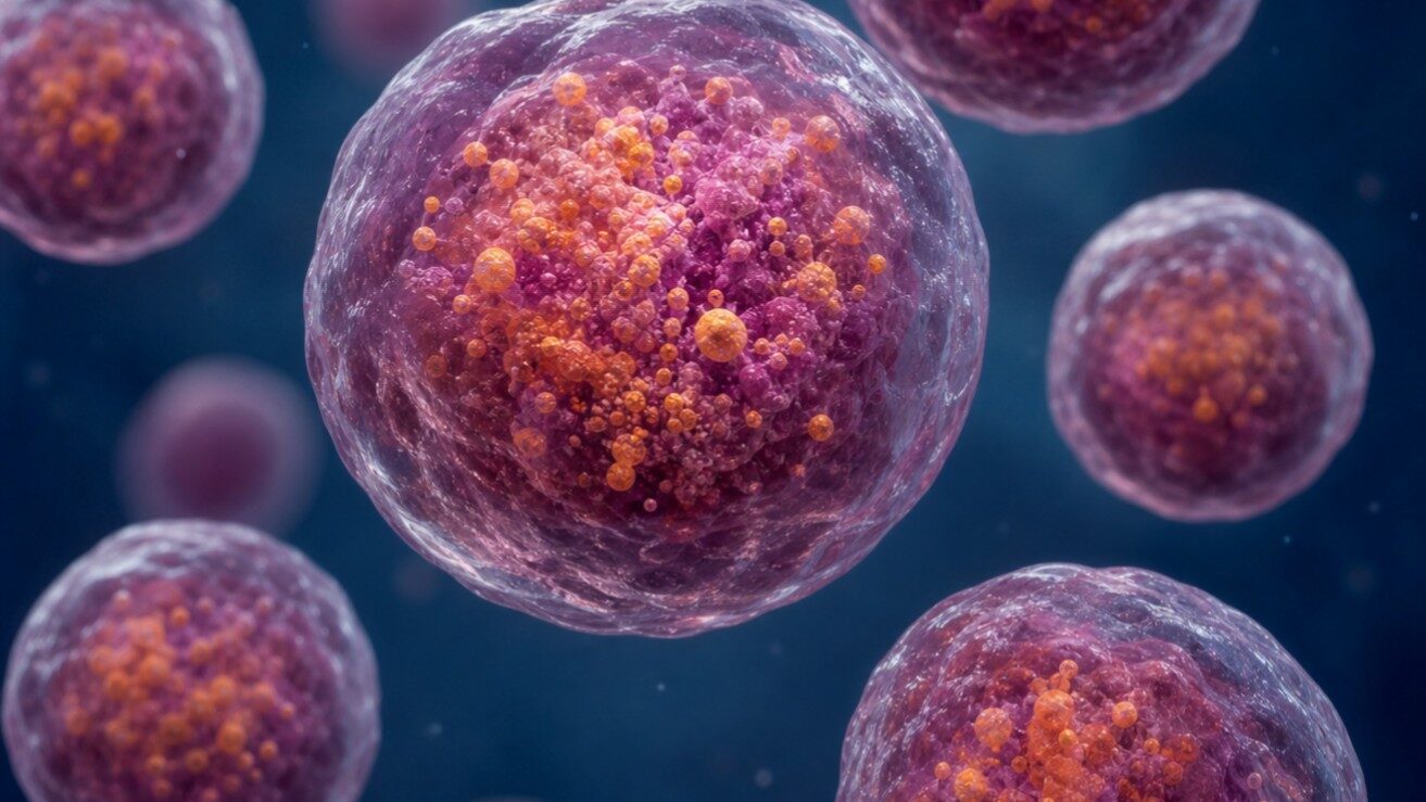
Your cells are constantly juggling two opposing needs: breaking things down and building things up. At the heart of that balancing act are lysosomes—tiny, acid-filled compartments that digest worn-out proteins, recycle cellular debris, and help cells decide whether it’s time to grow or conserve energy.
When lysosomes malfunction, the consequences can be serious. Lysosomal storage diseases, neurodegeneration, and metabolic disorders have all been linked to disrupted lysosome function. A new study published in Nature Communications has uncovered a key part of the control system that keeps lysosomes functioning properly.
Why Lysosome Location Matters
Lysosomes don’t just sit in one place. In well-fed cells, they spread out toward the cell’s edges, supporting active nutrient signaling and growth. But when nutrients run low, lysosomes cluster near the nucleus. This shift triggers elevated protein degradation and a cellular recycling process called autophagy.
A protein known as ARL8B plays a key role in driving lysosomes outward. When it’s active, lysosomes disperse. When it’s inactive, they cluster. But until now, no one had identified what turns ARL8B off.
Discovering the Brake
Researchers at the University of Bielefeld set out to find that missing brake. They identified a protein called TBC1D9B that directly binds active ARL8B and accelerates its inactivation. A membrane protein called TMEM55B anchors TBC1D9B to the lysosomal surface, positioning it to regulate ARL8B right where it’s needed. To confirm that TBC1D9B genuinely stimulates ARL8B inactivation, the researchers used the GTPase-Glo™ Assay to measure GTP hydrolysis directly. They found that wildtype TBC1D9B significantly accelerated ARL8B’s activity while a catalytically inactive mutant did not.
What Happens When the Brake Fails
Cells lacking TBC1D9B or TMEM55B showed lysosomes permanently dispersed to the cell periphery—unable to reposition in response to nutrient deprivation. When these cells were starved, lysosomal protease activity failed to increase and autophagic flux was significantly reduced.
Restoring TBC1D9B activity rescued normal lysosome behavior, confirming that its enzymatic function is essential. And when ARL8B was depleted alongside TBC1D9B loss, lysosomes re-clustered normally. This demonstrates that all of TBC1D9B’s effects depend on ARL8B.
A Newly Completed Circuit
These findings complete a regulatory circuit for lysosome positioning. One set of proteins activates ARL8B to disperse lysosomes when nutrients are plentiful. TMEM55B and TBC1D9B do the opposite: inactivating ARL8B to enable the perinuclear clustering cells need when nutrients run low.
This discovery matters beyond cell biology. Disrupted lysosome dynamics are linked to lysosomal storage diseases, Parkinson’s disease, cancer, and age-related decline. Understanding how lysosome dynamics are regulated at this molecular level opens new avenues for exploring disease mechanisms and, eventually, therapeutic intervention.
Reference: Duhay V., Tian M., Kosieradzka K. et al. (2026) Control of lysosome function by the GTPase-activating protein TBC1D9B and its binding partner TMEM55B. Nat. Commun. 17, 2487.