Bioluminescence and Biotechnology: Shining Nature’s Cool Light on Biology

Imagine you’re taking a refreshing night swim in the warm blue waters of Vieques in Puerto Rico. You splash into the surf and head out to some of the deeper waters of the bay, when what to your wondering eyes should appear, but blue streaks of light in water that once was clear. Do you need to get your eyes checked? Are you hallucinating? No! You’ve just happened upon a cluster of dinoflagellates, harmless bioluminescent microorganisms called plankton, that emit their glow when disturbed by movement. These dinoflagellates are known to inhabit waters throughout the world but are generally not present in large enough numbers to be noticed. There are only five ecosystems in the world where these special bioluminescent bays can be seen, and three of them are in Puerto Rico. 

Bioluminescent plankton exhibit a blue glow when disturbed.
Bioluminescent plankton in the ocean

But you don’t have to travel to Puerto Rico or swim with plankton to see bioluminescence. There are bioluminescent organisms all over the world in many unexpected places. There are bioluminescent mushrooms, bioluminescent sea creatures—both large and small (squid, jellyfish, and shrimp, in addition to the dinoflagellates)—and bioluminescent insects, to name a few. Bioluminescence is simply the ability of living things to produce light.

Continue reading “Bioluminescence and Biotechnology: Shining Nature’s Cool Light on Biology”
The Path Brightens for Vaccine Researchers: Luminescent Reporter Viruses Detect Neutralizing Antibodies

Developing a vaccine that is safe, effective, easily manufactured and distributed is a daunting task. Yet, that is exactly what is needed in response to the COVID-19 pandemic.

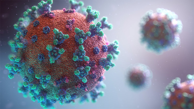
Computer generated 3D image of coronavirus

Vaccine development, safety and efficacy testing take time. The mumps vaccine is thought to be the quickest infectious disease vaccine ever produced, and its development required four years from sample collection to licensing (2). However, there are many reasons to anticipate quicker development for a COVID-19 vaccine: Researchers are collaborating in unprecedented ways, and most COVID-19 scientific publications are free for all to access and often available as preprints. As of August 11, 2020, researchers around the globe have more than 165 vaccine candidates in development, 30 of which are in some phase of human clinical trials (1). The range of vaccine formulations available to scientists has expanded to include RNA and DNA vaccines, replication-defective adenovirus vaccines, inactivated or killed vaccines and subunit protein vaccines. Equally important is that vaccine developers and researchers have greater access to powerful molecular biology tools like bioluminescent reporters that enable quicker testing and development.

Continue reading “The Path Brightens for Vaccine Researchers: Luminescent Reporter Viruses Detect Neutralizing Antibodies”

Choices for Measuring Luciferase-Tagged Reporter Pseudotyped Viral Particles in Coronavirus Research

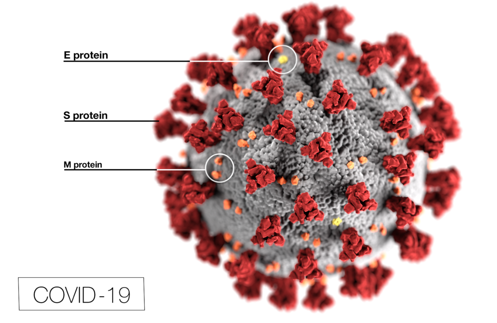
Coronavirus (CoV) researchers are working quickly to understand the entry of SARS-CoV-2 into cells. The Spike or S proteins on the surface of a CoV is trimer. The monomer is composed of an S1 and S2 domain. The division of S1 and S2 happens in the virus producing cell through a furin cleavage site between the two domains. The trimer binds to cell surface proteins. In the case of the SARS-CoV, the receptor is angiotensin converting enzyme 2. (ACE2). The MERS-CoV utilizes the cell-surface dipeptidyl peptidase IV protein. SARS-CoV-2 uses ACE2 as well. Internalized S protein goes though a second cleavage by a host cell protease, near the S1/S2 cleavage site called S2′, which leads to a drastic change in conformation thought to facilitate membrane fusion and entry of the virus into the cell (1).  

CDC / Alissa Eckert, MS; Dan Higgins, MAMS

Rather than work directly with the virus, researchers have chosen to make pseudotyped viral particles. Pseudotyped viral particles contain the envelope proteins of a well-known parent virus (e.g., vesicular stomatitis virus) with the native host cell binding protein (e.g., glycoprotein G) exchanged for the host cell binding protein (S protein) of the virus under investigation. The pseudotyped viral particle typically carries a reporter plasmid, most commonly firefly luciferase (FLuc), with the necessary genetic elements to be packaged in the particle. 

To create the pseudotyped viral particle, plasmids or RNA alone are transfected into cells and the pseudotyped viruses work their way through the endoplasmic reticulum and golgi to bud from the cells into the culture medium. The pseudoviruses are used to study the process of viral entry via the exchanged protein from the virus of interest. Entry is monitored through assay of the reporter. The reporter could be a luciferase or a fluorescent protein.   

Continue reading “Choices for Measuring Luciferase-Tagged Reporter Pseudotyped Viral Particles in Coronavirus Research”