Convenient, Non-Radioactive Detection of Isoaspartate

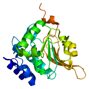
Structure of the PCMT1 protein. Based on PyMOL rendering of PDB 1i1n. Licensed under creative commons http://creativecommons.org/licenses/by-sa/3.0/deed.en

The ISOQUANT® Isoaspartate Detection Kit is intended for quantitative detection of isoaspartic acid residues in proteins and peptides, which can result from the gradual, nonenzymatic deamidation of asparagine or rearrangement of aspartic acid residues.

The ISOQUANT® Kit is designed to provide information regarding the global formation of isoaspartic acid residues at Asn and Asp sites, not at each site separately.

The deamidation of asparagine residues and rearrangement of aspartic acid residues is characterized by the formation of a succinimide intermediate that resolves to form a mixture of isoaspartic acid (typically 70–85%) and aspartic acid.
The rate and extent of isoaspartic acid formation can vary widely among proteins, depending on the amino acid sequence and size of the target protein. Deamidation of Asn residues has been observed most frequently at Asn-Gly and Asn-Ser sites within proteins.

The ISOQUANT® Isoaspartate Detection Kit uses the enzyme Protein Isoaspartyl ethyltransferase (PIMT) to specifically detect the presence of isoaspartic acid residues in a target protein. PIMT catalyzes the transfer of a methyl group from S-adenosyl-L-methionine (SAM) to isoaspartic acid. Spontaneous decomposition of this methylated intermediate results in the release of methanol and reformation of the succinimide.

References:

Wang, W. et al. (2012) Quantification and characterization of antibody deamidation by peptide mapping with mass spectrometry. Int. J. Mass. Spec. 312, 107–13.

Grappin, P. et al. (2011) New proteomic developments to analyze protein isomerization and their biological significance in plants. J. Proteomics, 74, 1475–82.

Yang, H. and Zubarev, R.A. (2010) Mass spectrometric analysis of asparagine deamidation and aspartate isomerization in polypeptides. Electrophoresis 31, 1764–71.

Sinha, S. et al. (2009) Effect of protein structure on deamidation rate in the Fc fragment of an IgG1 monoclonal antibody. Protein Sci. 18, 1573–84.
Endo H Application: Monitoring Protein Trafficking

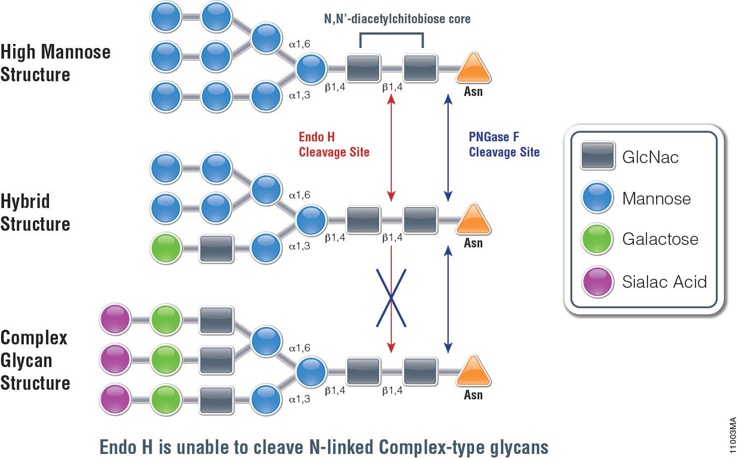
Endo H (Endo-ß-N-acetylglucosaminidase H) is a 29,000 dalton protein with optimal activity between pH 5 and 6. In contrast to PNGase F, which cleaves all N-linked glycans at the site of attachment to Asparagine (Asn), (with the exception of those with fucose attached to the core GlcNac moieties), Endo H hydrolyses the bond connecting the two GlcNac groups that comprise the chitobiose core (see Figure 1.). In addition, Endo H cleaves high mannose and hybrid glycans, whereas complex glycans (those with more than 4 different sugar types per glycan chain, including the GlcNac groups) are resistant to hydrolysis.

The unique specificty of Endo H and PNGase F can be used to monitor protein trafficking. Basic N-Glycosylation occurs in the endoplasmic reticulum. Proteins in this stage are sensitive to Endo H digestion. If proteins have entered the Golgi body where additional modifications occur to the glycan, they are resistant to Endo H digestion.

The following references illustrate this application:

Protein Profiling of a Lung Infection in a 500-Year-Old Mummy

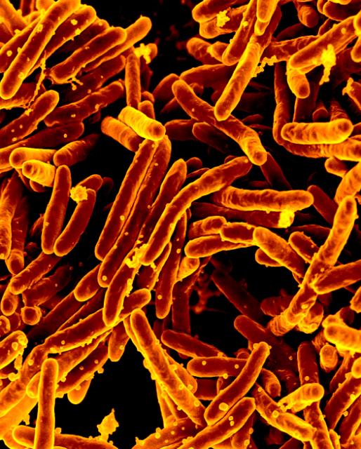
Mycobacterium tuberculosis Bacteria, the Cause of TB Scanning electron micrograph of Mycobacterium tuberculosis bacteria, which cause TB. Credit: NIAIDI am fascinated by all the ways that scientists are taking sensitive techniques and using them to look into our past. For example, scientists constructed the entire genome of Yersinia pestis, the caustive agent of the Black Death, from teeth and bone samples of plague victims from the 14th century. Without methods like polymerase chain reaction (PCR), such an analysis could not be performed. My fellow blogger Terri discussed how a postmortem autopsy of Ozti, a mummy found in the Alps, used modern techniques to learn not only what color his eyes were but that he suffered from Lyme disease. In a recent PLOS ONE article, Corthals et al. took this analysis of preserved human remains further to determine if a mummy from the Andes in Argentina may have suffered from an active lung infection, testing for an immune response by protein profiling. Continue reading “Protein Profiling of a Lung Infection in a 500-Year-Old Mummy”
Use of Nonspecific Proteases for Analysis of Proteins by Mass Spectrometry

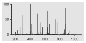
mass spectrometry results

One of the approaches to identify proteins by mass spectrometry includes the separation of proteins by gel electrophoresis or liquid chromatography. Subsequently the proteins are cleaved with sequence-specific endoproteases. Following digestion the generated peptides are investigated by determination of molecular masses or specific sequence. For protein identification the experimentally obtained masses/sequences are compared with theoretical masses/sequences compiled in various databases.

Are you looking for proteases to use in your research?
Explore our portfolio of proteases today.

Nonspecific proteases such as pepsin, proteinase K, elastase and thermolysin can offer an alternative to traditional sequence-specific proteases for certain applications. The following references illustrate the use of nonspecific proteinases for the mass spec analysis of proteins:

Papasotiriou, D. et al. (2010) Peptide mass fingerprinting after less specific in-gel proteolysis using MALDI-LTQ-Orbitrap and 4-chloro-alpha-cyanocinnamic acid. J. Proteome. Res. 9, 2619–29. This reference demonstrates the use of either chymotrypsin, elastase, trypsin or proteinase K in combination with matrix CHCA for increase peptide identification and sequence coverage using MALDI.

Neue, K. et al. (2011) Elucidation of glycoprotein structures by unspecific proteolysis and direct nanoESI mass spectrometric analysis of ZIC-HILIC-enriched glycopeptides. J. Proteome. Res. 10, 2248–60. Notes use of thermolysin or elastase in combination with ZIC-HILIC enrichment as alternative method for the characterization of glycopeptides.

Baeumlisberger, D et al. (2011) Simple dual-spotting procedure enhances nLC-MALDI MS/MS analysis of digests with less specific enzymes. J. Proteome. Res. 10, 2889–94. Data noted that samples digested with elastase followed by nLC separation and subsequent alternative spotting on both MALDI-LTQ-Orbitrap and MALDL-TOF/TOF instruments resulted in 32% additional peptides.

A New Method that Marks Proteins for Destruction

The ability to manipulate genes and proteins and observe the effects of specific changes is a foundational aspect of molecular biology. From the first site-directed mutagenesis systems to the development of knockout mice and RNA interference, technologies for making targeted changes to specific proteins to eliminate their expression or alter their function have made tremendous contributions to scientific discovery.
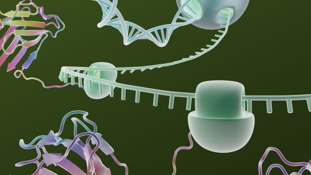
A recent paper highlights a novel application of HaloTag technology to enable the targeted destruction of specific HaloTag fusion proteins in vivo. The paper, published online in the July issue of Nature Chemical Biology, details a promising new method with application for validation of potential drug targets by specific in vivo inhibition, and for studying the function of specific genes in organogenesis or disease development. Continue reading “A New Method that Marks Proteins for Destruction”

Cell Free Expression Application: Production of Soluble Protein for Structural Analysis

The TNT® SP6 High-Yield Protein Expression System uses a high-yield wheat germ extract supplemented with SP6 RNA polymerase and other components. Coupling transcriptional and translational activities eliminates the inconvenience of separate in vitro transcription and purification steps for the mRNA, while maintaining the high levels of protein expression. All that is required is the addition of DNA templates containing the SP6 promoter and the protein coding region for the protein of interest. Furthermore no specialized equipment is required for protein screening and production. The system enables the expression of approximately 100µg/ml of protein in batch reaction and 200–440µg/ml in dialysis reaction in 10–20 hours .

In a recent publication (Zhao, L. et.al. (2010) J. Struct. Genomics 11, 201–9), the Northeast Structural Genomics Consortium (www.nesg.org) in their quest to express 5,000 eukaryotic proteins, report that even with different cloning strategies they could only produce 26% of the proteins in a soluble form. To improve the efficiency of expressing soluble protein, they investigated the use of wheat germ cell free system as a alternative to E.coli.

In this publication 59 human constructs were expressed in both E.coli and the wheat germ cell free system. Only 30% of human proteins could be produced in a soluble form using E.coli -based expression. Some 70% could be produced using the TNT® SP6 High Yield Wheat Germ system.
To further demonstrate the utility of expressing proteins that were suitable for structural studies from a wheat germ-based system, two of the proteins were isotope enriched and analyzed successfully by 2D NMR.

Use of Multiple Proteases for Improved Protein Digestion

One of the approaches to identify proteins by mass spectrometry includes the separation of proteins by gel electrophoresis or liquid chromatography. Subsequently the proteins are cleaved with sequence-specific endoproteases. Following digestion the generated peptides are investigated by determination of molecular masses or specific sequence. For protein identification the experimentally obtained masses/sequences are compared with theoretical masses/sequences compiled in various databases.

Trypsin is the favored enzyme for this application, for the following reasons: A) the peptides contain a basic residue (Arg or Lys) on the C terminus and thus are good candidates for collision induced activation (CAD) in tandem experiments (low charge states and high mass-to-charge ratios); B) it is relatively Inexpensive; and C) optimal digestion conditions have been well characterized.

An inherent limitation of trypsin is the size of the peptides that it generates. For most organisms > 50% of tryptic peptides are less than 6 amino acids, too small for mass spectrometry based sequencing.

Are you looking for proteases to use in your research?
Explore our portfolio of proteases today.

One recent publication examined the use of multiple proteases (trypsin, LysC, ArgC , AspN and GluC) in combination with either CAD or electron-based fragmentation (ETD) to improve protein identification (1). Their results indicated a significant improvement from a single protease digestion (trypsin), which yielded 27,822 unique peptides corresponding to 3313 proteins. In contrast using a combination of proteases with either CAD or ETD fragmentation methods yielded 92,095 unique peptides mapping to 3908 proteins.

Swaney DL, Wenger CD, & Coon JJ (2010). Value of using multiple proteases for large-scale mass spectrometry-based proteomics. Journal of proteome research, 9 (3), 1323-9 PMID: 20113005

Optimized Wheat Germ Extract for High-Yield Protein Expression of Functional, Soluble Protein

Wheat Germ Extract for high-yield protein expression

Cell-free protein synthesis has emerged as powerful alternative to cell based protein expression for functional and structural proteomics. The TNT® SP6 High-Yield Protein Expression System uses a high-yield wheat germ extract supplemented with SP6 RNA polymerase and other components. Coupling transcriptionaland translational activities eliminates the inconvenience of separate in vitro transcription and purification steps for the mRNA, while maintaining the high levels of protein expression (1).

Continue reading “Optimized Wheat Germ Extract for High-Yield Protein Expression of Functional, Soluble Protein”