Improved FFPE Tissue Sample Processing with High-Throughput Automated DNA Extraction

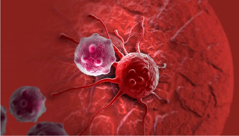
In oncology, tissue biopsies are commonly fixed in formalin and embedded in paraffin (FFPE). These FFPE samples can be used with immunohistochemical or molecular analysis for identifying biomarkers that guide the diagnosis and therapeutic management of patients. This fixation technique allows long-term storage of samples but impacts the integrity of nucleic acids. This makes extracting DNA and RNA from FFPE tissues in sufficient quantity and quality for molecular analysis techniques such as NGS analyses challenging for molecular oncology laboratories.

“At Rennes University Hospital, we receive many lung cancer samples with little material available, or samples of poor quality. The nucleic acid extraction step is therefore critical to get good yield. We have seen that it had a direct impact on the success of downstream analysis,” said Dr. Alexandra Lespagnol. Lespagnol is the Technical Manager of the Molecular Genetics of Cancer core lab at the University Hospital of Rennes in France.

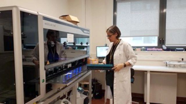
In order to accommodate the increasing number of samples that needed to be analyzed, the Molecular Genetics of Cancer core lab of the University Hospital of Rennes initiated an automation project for extracting DNA from FFPE tissues. The lab also wanted to improve sample tracking and reproducibility of their results.

Continue reading “Improved FFPE Tissue Sample Processing with High-Throughput Automated DNA Extraction”

RAS-Targeted Drug Discovery: From Challenge to Opportunity

cancer cell, ras-targeted drug discovery

In 1963, Jennifer Harvey was studying Moloney murine leukemia virus (MMLV) at the cancer research department of the London Hospital Research Laboratories. After routine transfers of plasma from MMLV-infected rats to mice, she made an unusual discovery. In addition to the expected leukemia, the mice that received the plasma developed solid tumors (soft-tissue sarcomas), primarily in the spleen (1). A few years later, Werner Kirsten at the University of Chicago observed similar results working with mouse erythroblastosis virus (MEV) (2).

Subsequent research, with the advent of genome sequencing, showed that a cellular rat gene had been incorporated into the viral genome in both cases (3). These genomic sequences contained a mutation later shown to be responsible for the development of sarcomas, and the word “oncogene” became a common part of the vocabulary in cancer publications during the early 1980s (4). Harvey’s discovery led to the naming of the corresponding rat sarcoma oncogene as HRAS, while Kirsten’s related oncogene was named KRAS. Several laboratories, working independently, cloned the human homolog of the viral HRAS gene in 1982 (3). The human KRAS gene was cloned shortly thereafter, as well as a third RAS gene, named NRAS (3). Additional studies showed that a single point mutation in each of these genes led to oncogenic activation, and they have been popular targets for anticancer drug discovery efforts ever since.

Continue reading “RAS-Targeted Drug Discovery: From Challenge to Opportunity”

When Good Proteins Go Bad

Ribbon model of p53 protein bound to DNA molecule.

Following what feels like an exceptionally long and brutal winter, I for one couldn’t be happier about the arrival of Spring and the way it makes everything seem brighter and brand-new. Soaking in the soul-warming sunshine. Reveling in the sweet melody of chirping birds. Watching the earth literally coming alive again with greenery. And for those of us who love and are enthralled by scientific discoveries like myself, the report of a recent shiny new discovery in the world of cancer research is equally as day-brightening and spirit-lifting.

To suppress tumors or to not suppress tumors: that is the question.

In the world of oncology, the protein known as p53 has long proven itself to be a primary target of interest. p53 operates as a tumor suppressor protein, often lauded as the “guardian of the human genome”, due to its dedication to governing controlled cell division and assessing damaged DNA. There are a number of cellular stressors that can wreak havoc on your DNA, including exposure to ultraviolet light or radiation, oxygen deficiency (hypoxia), and contact with hazardous chemicals.

Consider a normal-functioning p53 protein as the quality control person in a production factory. The p53 protein evaluates the products, DNA, coming down the line and determines an appropriate course of action for those that do not meet the quality standards.

Let’s say some less-than-quality DNA comes down the pipe. If the DNA is not too severely injured, p53 will alert and activate additional genes to repair the damage. However, if the products coming through are too marred to repair, p53 will shut down the whole factory, if you will, by signaling for the cell to self-destruct via apoptosis. In doing so, p53 effectively impedes tumor development by inhibiting the ability for flawed DNA to further divide.

So, it would seem like p53 has proven itself to be an undeniably upstanding citizen of the protein variety, right? The unfortunate truth of the matter is p53 balances delicately on a double-edged sword, establishing itself as the veritable Dr. Jekyll and Mr. Hyde of the cellular world: usually unquestionably good, but sometimes unspeakably evil. Continue reading “When Good Proteins Go Bad”

Shining a Bright Light on Deep Questions in Biology with Bioluminescence

artists view inside a cell

Search the PubMed database for “dual-luciferase” and citations abound. The Dual-Luciferase® Reporter Assay is a powerful tool that allows researchers to ask a multitude of questions about gene control and expression in a system that itself could be normalized and internally controlled. For more than 15 years, firefly and Renilla luciferases  have formed the basis of a range of powerful assays and research tools for scientists who are asking questions about the deep and complex genetic and cellular story associated with cancer. Here we talk a bit of about bioluminescent chemistries, some of the newest bioluminescent tools available, and how some of these tools can be used to probe the deeper questions of cell biology, including cancer biology.

Continue reading “Shining a Bright Light on Deep Questions in Biology with Bioluminescence”