RIPK1: Promising Drug Target of Chronic Inflammatory Diseases

Today’s post is written by Michael Curtin, Senior Product Manager, Reporters and Signaling.

Inflammation is a defense mechanism that the body employs in which the immune system recognizes and removes harmful and foreign stimuli and begins the healing process. Inflammation can be either acute or chronic. Chronic inflammation is also referred to as slow, long-term inflammation and can last for prolonged periods (several months to years); chronic inflammation is caused by immune dysregulation. This typically takes the form of the body’s inability to resolve inflammation resulting from overproduction of inflammatory cytokines and chemokines, as well as danger-associated molecular patterns (DAMPs) released from dying cells (2). Tumor Necrosis Factor (TNF) is the primary cytokine involved in many common inflammatory diseases and is where many therapies targeting inflammation are focused.
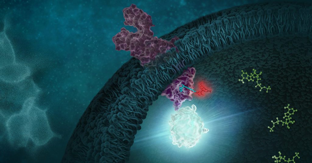
Signaling of kinases like RIPK1 can be studied using the NanoBRET target engagement assays

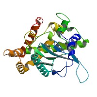
Recent research that RIP kinases (RIPK1 and RIPK3) are important regulators of innate immunity via their key roles in cell death signaling during cellular stress and following exposure to inflammatory and infectious stimuli. RIPK1 has an important scaffolding role in pro-inflammatory signaling where it interacts with TRADD, TRAF1 TRAF2, and TRAF3 and TRADD can act as an adaptor protein to recruit RIPK1 to the TNFR1 complex in a TNF-dependent process. RIPK1 plays a kinase activity-dependent role in both apoptotic and necroptotic cell death. A review article by Speir et al. (1) discusses the role of RIP kinases in chronic inflammation and the potential of RIPK1 inhibitors as a new therapeutic approach for the treatment of chronic inflammation. RIPK1 or Receptor Interacting Protein Kinase 1 is a serine/threonine kinase that was originally identified as interacting with the cytoplasmic domain of FAS. Promega offers several reagents that make studying RIPK1 easier- these include our RIPK1 Kinase Enzyme Systems which includes RIPK1 (Human, recombinant; amino acids 1-327), myelin basic protein (MBP) substrate, reaction buffer, MnCl2, and DTT and is optimized for use with our ADP-Glo Kinase Assay.

Continue reading “RIPK1: Promising Drug Target of Chronic Inflammatory Diseases”

Increasing Drug Research and Development Efficiency Using a 4-point Screening Method to Determine Molecular Mechanism of Action

Fig 4. Four point MMOA screen for tideglusib and GW8510. Time dependent inhibition was evaluated by preincubation of TbGSK3β with 60 nM tideglusib and 6 nM GW-8510 with 10μM and 100μM ATP. (A). Tideglusib [60 nM] in 10μM ATP. (B). GW8510 [60 nM] in 10μM ATP. (C.) Tideglusib [60 nM] at 100μM ATP. (D.) GW8510 [60 nM] at 100μM ATP. All reactions preincubated or not preincubated with TbGSK3β for 30 min at room temperature. Experiments run with 10μM GSM peptide, 10μM ATP, and buffer. Minute preincubation (30 min) was preincubated with inhibitor, TbGSK3β, GSM peptide, and buffer. ATP was mixed to initiate reaction. No preincubation contained inhibitor, GSM peptide, ATP, and buffer. The reaction was initiated with TbGSK3β. Reactions were run at room temperature for 5 min and stopped at 80°C. ADP formed was measured by ADP-Glo kit. Values are mean +/- standard error. N = 3 for each experiment and experiments were run in duplicates. Control reactions contained DMSO and background was determined using a zero time incubation and subtracted from all reactions. Black = 30 min preincubation Grey = No preincubation.
Four point MMOA screen for tideglusib and GW8510.
Time dependent inhibition was evaluated by preincubation of TbGSK3β with 60 nM tideglusib and 6 nM GW-8510 with 10μM and 100μM ATP. (A). Tideglusib [60 nM] in 10μM ATP. (B). GW8510 [60 nM] in 10μM ATP. (C.) Tideglusib [60 nM] at 100μM ATP. (D.) GW8510 [60 nM] at 100μM ATP. All reactions preincubated or not preincubated with TbGSK3β for 30 min at room temperature.  Black = 30 min preincubation Grey = No preincubation.
The first small-molecule kinase inhibitor approved as a cancer therapeutic, imatinib mesylate (Gleevec® treatment), has been amazingly successful. However, a thorough understanding of its molecular mechanism of action (MMOA) was not truly obtained until more than ten years after the molecule had been identified.

Understanding the MMOA for a small-molecule inhibitor can play a major role in optimizing a drug’s development. The way a drug actually works–the kinetics of binding to the target molecule and how it competes with endogenous substrates of that target–ultimately determines whether or not a a candidate therapeutic can be useful in the clinic. Drugs that fail late in development are extremely costly.

Drug research and discovery for neglected tropical diseases suffer from a lack of a large commercial market to absorb the costs of late-stage drug development failures. It becomes very important to know as much as possible, simply and quickly, about MMOA for candidate molecules for these diseases that are devastating to large populations.

One such neglected topical disease is Human African trypanosomiasis (HAT, also known as sleeping sickness). Continue reading “Increasing Drug Research and Development Efficiency Using a 4-point Screening Method to Determine Molecular Mechanism of Action”

Identifying and Profiling Inhibitors for PI 4-Kinases Using a Luminescent High-Throughput Screen

The phosphotidylinositol 4-kinases (PI 4-kinases) generate phosphotidyl-4-phosphate (PI(4)P) from phosphotylinositol. PI(4)P is an important precursor for other phosphoinositides involved in signaling, such as PI(4,5)P2, which is the substrate of phospholipase C (PLC) and the precursor of DAG and insitol (1,4,5) triphosphate.

There are four different mammalian PI 4-kinases currently described, and these have been divided into two classes based on their sensitivities to wortmannin and adenosine. Type II PI 4-kinases (PI4K2A and PI4K2B) are not sensitive to wortmannin, but are inhibited by the nonspecific inhibitor adenosine; Type III PI 4-kinases (PI4KA and PI4KB) are sensitive to wortmannin.

The functions of the PI 4-kinases and their products are not fully understood. At least one study has shown that PI 4-kinases are important for the proper recycling of synaptic vesicles. The PI 4-kinase from Drosophila, four-wheel drive, is critical for contractile ring formation during cytokinesis. Other studies in yeasts and mammals have shown that PI 4-kinases localize to the Golgi, and in mammals might be critical for proper budding of vesicles from the Golgi. Additionally, type III PI 4-kinases appear to play a role in the replication of hepatitis C virus (HCV) and enteroviruses by participating in the formation of altered host membrane structures. Although, we have hints about their function, to really understand and dissect the precise roles of PI 4-kinases in cells, new tools, such as specific small-molecule inhibitors are required.

Continue reading “Identifying and Profiling Inhibitors for PI 4-Kinases Using a Luminescent High-Throughput Screen”

A Sensitive, Universal Kinase Assay Ideal for Use with Low-Turnover Enzymes

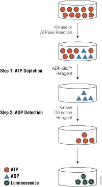
ADP-Glo™ Kinase Assay flowchart

Promega carries a large array of luminescent-based assays to measure cellular events such as viability, cytochrome P450 activity and apoptosis. Recently, we launched a new universal, homogeneous, high-throughput screening method called the ADP-Glo™ Kinase Assay, which measures kinase activity by quantifying the amount of ADP produced during a kinase reaction. While we already offer the Kinase-Glo™ Assays for assessing the quantity of ATP remaining after a kinase reaction, these assays are not ideal for use with low-activity kinases.

Continue reading “A Sensitive, Universal Kinase Assay Ideal for Use with Low-Turnover Enzymes”