New Uses for Old Drugs: Remdesivir and COVID-19

With the COVID-19 pandemic far from over in the United States and worldwide, the battle against the disease continues to intensify. Much hope has been pinned on vaccine development. However, vaccines are a long-term, preventative strategy. The immediate need for drugs to fight COVID-19 has accelerated efforts for a variety of potential treatments (see The Race to Develop New Therapeutics Against Coronaviruses).

The Remdesivir Origin Story

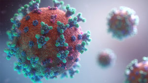
3d model of coronavirus

One drug that has received widespread attention is remdesivir. It was developed from research by Gilead Sciences that began in 2009, originally targeting hepatitis C virus (HCV) and respiratory syncytial virus (RSV) (1). At present, remdesivir is classified as an investigational new drug (IND) and has not been approved for therapeutic use anywhere in the world.

Continue reading “New Uses for Old Drugs: Remdesivir and COVID-19”

How the SARS-CoV-2 Coronavirus Enters Host Cells and How To Block It

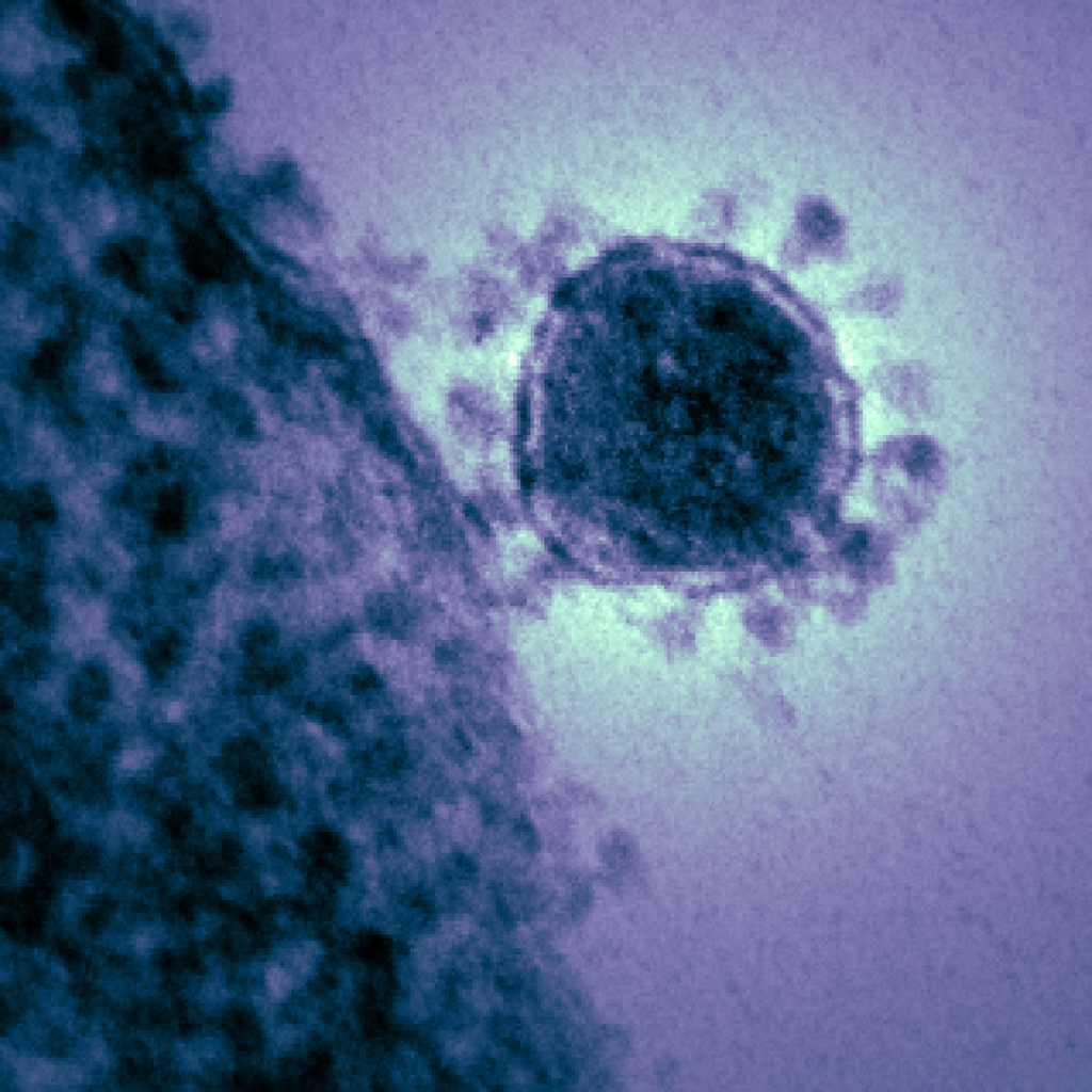
TE micrograph of a single MERS-CoV
Photo courtesy of National Institute of Allergy and Infectious Diseases

In December 2019, a new disease emerged from a seafood market in Wuhan, China. People who were infected began experiencing fever, dry cough, muscle aches and shortness of breath. The disease swept through China like wildfire and quickly spread overseas to almost every continent. We now know the virus that caused this disease, SARS-CoV-2, is a member of the severe acute respiratory syndrome coronavirus, and the disease itself was officially named COVID-19. According to the Johns Hopkins University Coronavirus Resource Center, there are 877,422 confirmed cases of COVID-19 worldwide, and 43,537 total deaths at the publication of this blog. Those numbers are only expected to increase over the next few weeks.

In this moment of crisis, scientists all around the world are desperately trying to find ways to treat and prevent the disease. One strategy for preventing the spread of the virus is to block its entry into human cells. But first we need to understand how SARS-CoV-2 enters human cells. A research group at the German Primate Center led by Dr. Stefan Pohlmann provides some answers in a recent publication in Cell.

Continue reading “How the SARS-CoV-2 Coronavirus Enters Host Cells and How To Block It”

Investigation of Remdesivir as a Possible Treatment for SARS-2-CoV (2019-nCoV)

Remdesivir (RDV or GS-5734) was used in the treatment of the first case of the SARS-CoV-2 (formerly 2019-nCoV ) in the United States (1). RDV is not an approved drug in any country but has been requested by a number of agencies worldwide to help combat the SARS-CoV-2 virus (2). RDV is an adenine nucleotide monophosphate analog demonstrated to inhibit Ebola virus replication (3). RDV is bioactivated to the triphosphate form within cells and acts as an alternative substrate for the replication-necessary RNA dependent RNA polymerase (RdRp). Incorporation of the analog results in early termination of the primer extension product resulting in the inhibition.

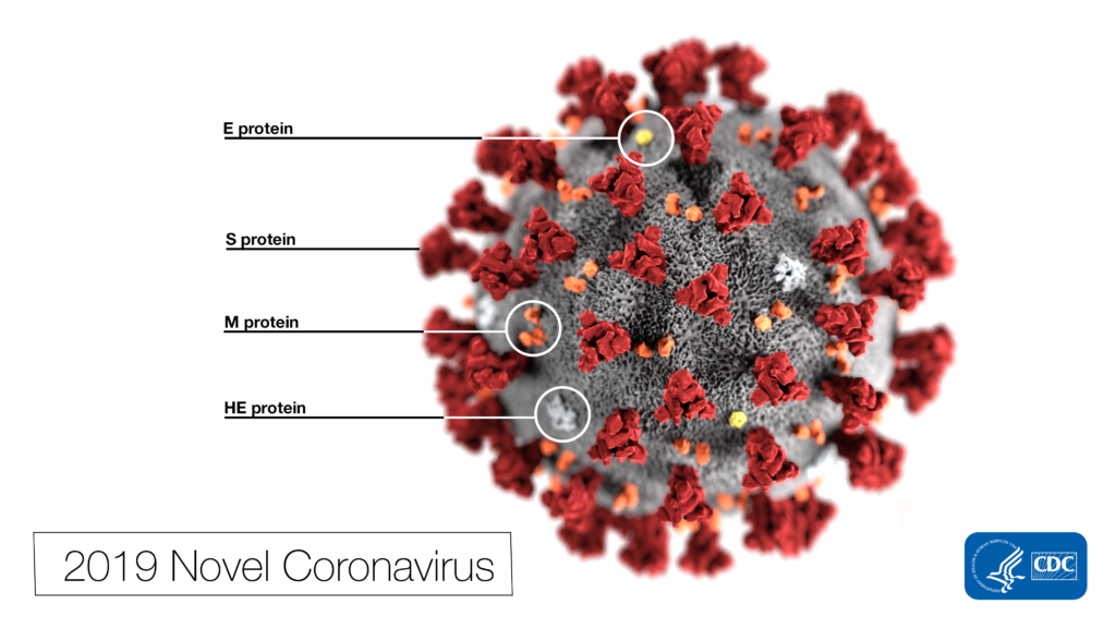
 Note the spikes that adorn the outer surface of the virus, which impart the look of a corona surrounding the virion, when viewed electron microscopically. In this view, the protein particles E, S, M, and HE, also located on the outer surface of the particle, have all been labeled as well. A novel coronavirus virus was identified as the cause of an outbreak of respiratory illness first detected in Wuhan, China in 2019.
This illustration, created at the Centers for Disease Control and Prevention (CDC), reveals ultrastructural morphology exhibited by coronaviruses. Photo Credit: Alissa Eckert, MS; Dan Higgins, MAM CDC

Why all the interest in RDV as a treatment for SARS-CoV-2 ? Much of the interest in RDV is due to a series of studies performed by collaborating groups at the University of North Carolina Chapel Hill (Ralph S. Baric’s lab) and Vanderbilit University Medical Center (Mark R. Denison’s lab) in collaboration with Gilead Sciences. 

Continue reading “Investigation of Remdesivir as a Possible Treatment for SARS-2-CoV (2019-nCoV)”

Using CellTiter-Glo® Luminescent Cell Viability Assay to Assess Cell Viability in Cancer Cells Treated with Silver Nanoparticles and DNA-PKcs Inhibitor

Silver nanoparticles (Ag-np) are commonly used in many consumer products, including cosmetics, textiles, electronics and medicine, largely due to their antimicrobial properties. More recently, Ag-np are being used to target and kill cancer cells. It has been known for years that silver nanoparticles (Ag-np) can induce cell death and DNA damage. Studies have also shown that Ag-np inhibit cell proliferation and induce apoptosis in cancer cells. However, cancer cells are able to fight back with DNA repair mechanisms such as non-homologous end joining repair (NHEJ). The NHEJ pathway requires the activation of DNA-dependent protein kinase catalytic subunit (DNA-PKcs), thus DNA-PKcs may protect against the Ag-np-induced DNA damage in cancer cells.

Could inhibition of DNA-PKcs increase the ability of Ag-np to kill cancer cells? In a 2017 study, Lim et al. wanted to test whether inhibition of DNA-PKcs can increase the cytotoxic effect of Ag-np in breast cancer and glioblastoma cell lines. To effectively determine cell viability in these cancer cell lines, the authors used the CellTiter-Glo® Luminescent Cell Viability Assay. The CellTiter-Glo® Assay determines the number of viable cells in culture based on quantitation of ATP, an indicator of metabolically active cells. A major advantage of this assay is its simplicity. This plate-based assay involves adding the single reagent (CellTiter-Glo® Reagent) directly to cells cultured in serum-supplemented medium. This generates a luminescent signal proportional to the amount of ATP present, which is detected using a luminometer. Cell washing, removal of medium and multiple pipetting steps are not required. Another advantage of the CellTiter-Glo® Assay is its high sensitivity. The system detects as few as 15 cells/well in a 384-well format in 10 minutes after adding reagent and mixing, making it ideal for automated high-throughput screening, cell proliferation and cytotoxicity assays.

The authors first confirmed that Ag-np treatment reduced proliferation and induced cell death/DNA damage in two breast cancer cell lines and two glioblastoma cell lines. The cytotoxic effect of Ag-np is specific to cancer cells, as minimal cytotoxicity was observed in non-cancerous human lung fibroblasts used as control. Next, they pre-treated the cancer cells with a DNA-PKcs inhibitor for 1 hour before adding Ag-np. Inhibition of DNA-PKcs increased Ag-np-mediated cell death in all four cancer cell lines. This suggests that DNA-PKcs may be protecting the cells from Ag-np cytotoxicity. The authors further showed that DNA-PKcs may repair Ag-np induced DNA damage by NHEJ and JNK1 pathways. In addition, DNA-PKcs may help recruit DNA repair machinery to damaged telomeres.

This study suggests that a combination of Ag-np treatment and DNA-PKcs inhibition may be a potential strategy to enhance the anticancer effect of Ag-np.

Reference: Hande M.P., et.al. (2017) DNA-dependent protein kinase modulates the anti-cancer properties of silver nanoparticles in human cancer cells. Mutat Res Gen Tox En. 824, 32

Screening for Inhibitors of CD73 (5´-ectonucleotidase) Using a Metabolite Assay

CD73CD73 also known as 5´-Ectonucleotidase (NT5E) is a membrane-anchored protein that acts at the outer surface of the cell to convert AMP to adenosine and free phosphate. CD73 activity is associated with immunosuppression and prometastatic effects, including angiogenesis. CD73 is highly expressed on the surfaces of many types of cancer cells and other immunosuppressive cells (1). A recent study by Quezada and colleagues showed that the high concentration of adenosine produced by the CD73-catalyzed reaction on glioblastoma multiforme cells, which are characterized by extreme chemoresistance, triggered adenosine signaling and in turn, the multi-drug resistance (MDR) phenotype of these cells (2).

Because of the roles of adenosine in immunosuppression, angiogenesis and MDR phenotypes, CD73 (NT5E) is an attractive therapeutic target. However, the current methods of assaying for the ectonucleotidase activity, HPLC and a malachite green assay, are cumbersome and not suited to high-throughput screening. The HPLC assay is expensive and difficult to automate and miniaturize (3). The malachite green assay is sensitive to phosphate found in media, buffers and other solutions used in the compound-screening environment.

To address the problem of developing a reliable high-throughput screening assay for CD73, Sachsenmeier and colleagues (3) looked to a luminescent ATP-detection reagent. Continue reading “Screening for Inhibitors of CD73 (5´-ectonucleotidase) Using a Metabolite Assay”