Looking Back: Cell-Free Expression Systems Helped to Characterize Proteins Involved in Hypoxia Response

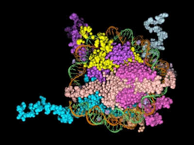
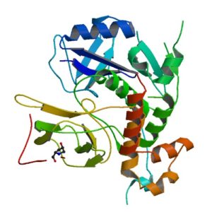
Structur of a HIF-1a-pVHL-ElonginB-ElonginC complex
Structure of a HIF-1a-pVHL-ElonginB-ElonginC complex

William G. Kaelin Jr., Sir Peter J. Ratcliffe and Gregg L. Semenza were awarded the 2019 Nobel Prize in Physiology or Medicine for their discoveries of how cells sense and adapt to oxygen availability.

Kaelin and Ratcliffe’s labs focused their efforts on the transcription factor HIF (hypoxia-inducible factor). This transcription factor is critical in the cellular adaptation of to changes in oxygen availability.

When oxygen levels are elevated cells contain very little HIF. Ubiquitin is added to the HIF protein via the VHL complex and it is degraded in the proteasome.  When oxygen levels are low (hypoxia) the amount of HIF increases.

In 2001 both groups published articles characterizing the interaction between VHL and HIF, and these articles were referenced by the Nobel Prize Organization in their press release about this year’s award. (1,2). Both studies demonstrated that under the normal oxygen conditions hydroxylation of proline residue P564 enabled VHL to recognize and bind to HIF.

The use of cell free expression (i.e., TNT Coupled Transcription/Translation System) by both labs was key in the characterization of the VHL:HIF interaction The labs utilized HIF and VHL 35-S labeled proteins generated via the TNT system under both normal or in a hypoxic work station to:

  • Determine the affect of ferrous chloride and cobaltous chloride on the interaction
  • Map the specific region of HIF required for the interaction to occur (556-574)
  • Determine the effect of HIF point mutations on the interaction
  • Use synthetic peptides to block the interaction
  • Conclude that a factor in mammalian cells was necessary for the interaction to occur.

Literature Cited

  1. Ivan, M et al. (2001) HIF Targeted for VHL-Mediated Destruction by Proline Hydroxylation: Implications for O2 Sensing. Science 292: 464–67.
  2. Jaakkola, P. et al. (2001) Targeting of HIF-α to the von Hippel-Lindau Ubiquitylation of Complex by O2– Regulated Prolyl Hydroxylation. Science 202, 468–72 .
Related Posts

Moving Out of the Cell: Advantages of Cell-Free Protein Expression

Cell-free protein expression is a simplified and accelerated avenue for the transcription and/or translation of a specific protein in a quasi cell environment. An alternative to slower, more cumbersome cell-based methods, cell-free protein expression methods are simple and fast and can overcome toxicity and solubility issues sometimes experienced in the traditional E. coli expression systems.

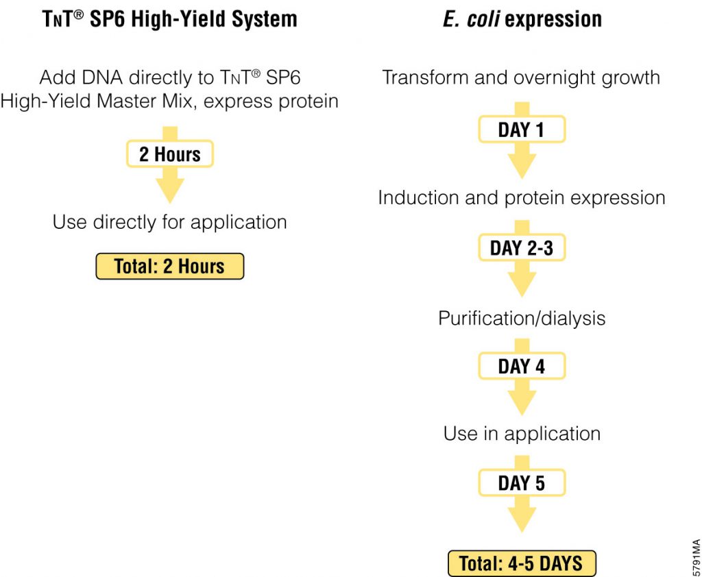
Cell-free protein expression offers significant time savings over cell-based expression methods.

Cell-free protein expression offers a convenient method for generating small amounts of protein for a variety of applications (e.g., protein:protein interactions, protein: nucleic acid interactions, structural analysis, functional assays and toxicity screening). This approach lends itself to specific protein labeling with fluorescence, biotin, radioactivity or heavy atoms, via modified charged tRNA’s or amino acids. Cell-free protein expression systems provide quick access to proteins of interest and remain a staple in the collection of tools available for the elucidation of protein structure and function, understanding cellular pathways and mechanisms and high-throughput screening of compounds for drug discovery. There are a number of different cell-free expressions systems, each with different strengths. Deciding which one is right for you depends upon your research needs and goals.

Continue reading “Moving Out of the Cell: Advantages of Cell-Free Protein Expression”

Cell-free expression application: Screening for successful oligo-mediated knockdown design

800px-ZebrafischAlthough previous references have provided data regarding the potential oncogenic role of the gene ETV7, there has been minimal investigation as to its physiological role.
In the following reference, Quintana, A. et al. (2014) Disease Models & Mechanisms 7, 265–70, zebrafish were used as in vivo model system to characterize ETV7.

One key experiment required the morpholino-oligonucleotide -mediated knockdown of in vivo ETV7. Two independent morpholinos were designed: one that inhibited translation and the other that inhibited proper splicing of exon 3. The efficacy of the translation –blocking morpholino was assessed with cell free expression of ETV7-tagged with hemagglutinin (HA).

Western blot performed with anti-HA antibodies determined the extent of the knockdown compared to a control containing no morpholino added. Once an efficient design was determined via cell-free expression screening, it was used for in vivo experiments. In conjunction additional other techniques, concluded that ETV7 is essential for normal red blood cell development.

Characterization of Ubiquitination Using Cell-Free Expression

Ubiquitination refers to the post translational modification of a protein by attachment of one or more ubiquitin monomers. The most prominent function of ubiqutin is labeling proteins for proteasome degradation. In addition to this function ubiquitination also controls the stability, function and intracellular localization of a wide variety of proteins.

Cell free expression can be used to characterize ubiquitation of proteins. Target proteins are expressed in a rabbit reticulocyte cell free system (supplemented with E1 ubiquitin activating enzyme, E2 ubiquitin –conjugating enzyme, and ubiquitin). Proteins that have been modified can be analyzed by a shift in migration on polyacrylamide gels.

The following references illustrate the use of cell free expression for this application.

Jung, Y.S. et al. (2011) The p73 Tumor Suppressor Is Targeted by Pirh2 RING Finger E3 Ubiquitin Ligase for the Proteasome-dependent Degradation. J. Biol. Chem. 286, 35388–95.

Su, C-H, et al. (2010) 14-3-3sigma exerts tumor-suppressor activity mediated by regulation of COP1 stability. Cancer. Res. 71, 884–94.

Naoe, H. et al. (2010). The anaphase-promoting complex/cyclosome activator Cdh1 modulates Rho GTPase by targeting p190 RhoGAP for degradation. Mol. Cell. Biol. 30, 3994-05.

de Thonel, A. et al. (2010) HSP27 controls GATA-1 protein level during erythroid cell differentiation. Blood 116, 85–96.

Kaneko, M. et al. (2010) Loss of HRD1-mediated protein degradation causes amyloid precursor protein accumulation and amyloid-beta generation. J. Neurosci. 30, 3924–32.

Optimized Protein Expression: Flexi Rabbit Reticulocyte Lysate

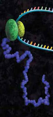
A protein chain being produced from a ribosome.

mRNAs commonly exhibit differing salt requirements for optimal translation. Small variations in salt concentration can lead to dramatic differences in translation efficiency. The Flexi® Rabbit Reticulocyte Lysate System allows translation reactions to be optimized for a wide range of parameters, including
Mg2+ and K+ concentrations and the choice of adding DTT. To help optimize Mg2+ for a specific message, the endogenous Mg2+ concentration of each lysate batch is stated in the product information included with this product.

The following references utilize the features of Flexi Rabbit Reticulocyte Lysate System to investigate certain parameters of translation:

Vallejos, M. et al. (2010)The 5′-untranslated region of the mouse mammary tumor virus mRNA exhibits cap-independent translation initiation. Nucl Acids Res. 38, 618–32. Identification of internal ribosomal ribosomal entry site in the 5’ untranslated region of the mouse mammary tumor virus mRNA.

Spriggs, K. et al. (2009) The human insulin receptor mRNA contains a functional internal ribosome entry segment. Nucl. Acids. Res. 17, 5881–93. Identification of a functional internal ribosome entry site in the human insulin receptor mRNA.

Powell, M. et al. (2008) Characterization of the termination-reinitiation strategy employed in the expression of influenza B virus BM2 protein. RNA 14, 2394–06. Analysis of the mRNA signals involved in the expression of influenza B virus BM2 protein.

Sato, V. et al. (2007) Measles virus N protein inhibits host translation by binding to eIF3-p40. J. Vir. 81, 11569–76. Charaterized the effect of the measles virus N protein binding to the translation initiation factor eIF3-p40 on the expression of various proteins in rabbit reticulocyte lysate.

Hirao, K. et al. (2006) EDEM3, a soluble EDEM homolog, enhances glycoprotein endoplasmic reticulum-associated degradation and mannose trimming. J. Biol. Chem. 281, 9650–58. The EDEM3 protein was expressed in the presence of canine microsomal membranes to establish that co-translational translocation occurs into the endoplasmic reticulum.

Shenvi, C. et al. (2005) Accessibility of 18S rRNA in human 40S subunits and 80S ribosomes at physiological magnesium ion concentrations–implications for the study of ribosome dynamics. RNA 11, 1898–08. Characterization of ribosome dynamics under different ionic conditions.

Nair, A. et al. (2005) Regulation of luteinizing hormone receptor expression: evidence of translational suppression in vitro by a hormonally regulated mRNA-binding protein and its endogenous association with luteinizing hormone receptor mRNA in the ovary. J. Biol. Chem. 280, 42809–16. Examined the affect of luteinizing hormone receptor mRNA binding protein on transltional suppression of luteinizing hormone receptor RNA.

Use of Cell-Free Protein Expression for Epigenetics-Related Applications

Epigenetics is the study of the processes involved in the genetic development of an organism, especially the activation and deactivation of genes. One way that genes are regulated is through the remodeling of chromatin. Chromatin is the complex of DNA and the histone proteins with which it associates. The conformation of chromatin is profoundly influenced by the post-translational modification of the histone proteins. These modifications include acetylation, methylation, ubiquitylation, phosphorylation and sumolyation. The following references illustrate the use of cell-free expression to characterize this process.

Shao, Y. et al. (2010) Nucl. Acid. Res. 38, 2813–24.
Carbonic anhydrase IX (CAIX) plays an important role in the growth and survival of tumor cells.The MORC proteins contain a CW-type zinc finger domain and are predicted to have the function of regulating transcription, but no MORC2 target genes have been identified. CAIX mRNA to be down-regulated 8-fold when MORC2 was overexpressed. Moreover, MORC2 decreased the acetylation level of histone H3 at the CAIX promoter. Among the six HDACs tested, histone deacetylase 4 (HDAC4) had a much more prominent effect on CAIX repression. Assays showed that MORC2 and HDAC4 were assembled on the same region of the CAIX promoter. Interaction between MORC2 and HDAC 4 were confirmed by using cell free expression of MORC2 and GST-HDAC (GST pull-downs). Cell-free expression was also used to express MORC2 proteins to determine through gel shifts the binding location on the CAIX promoter region (gel shift experiments)

Denis, H. et al. (2009) Mol. Cell. Biol. 29, 4982–93.
The recent identification of enzymes that antagonize or remove histone methylation offers new opportunities to appreciate histone methylation plasticity in the regulation of epigenetic pathways. PAD4 was the first enzyme shown to antagonize histone methylation. Very little is known as to how PADI4 silences gene expression. Through the use of cell-free expression to express both PAD4 and HDAC1 proteins and E. coli expression of GST fusions of PAD4 and HDAC1, pulldown experiments confirmed by in vivo experiments that PADI4 associates with the histone deacetylase 1 (HDAC1), and the corresponding activities, associate cyclically and coordinately with the pS2 promoter during repression phases.

Brackertz, M. et al. (2006) Nucl. Acid. Res. 34, 397-406.
The Mi-2/NuRD complex is a multi-subunit protein complex with enzymatic activities involving chromatin remodeling and histone deacetylation. The function of p66α and of p66β within the multiple subunits has not been addressed. GST-fused histone tails of H2A, H2B, H3 and H4 were expressed in E. coli used in an in vitro pull-down assay with radioactively labeled p66-constructs expressed using cell free systems. Deletions at the C terminus noted reduced binding of p66 where as deletions at the N terminus did not affect binding. Also observed was that acetylation of histone tails reduces the association with both p66-proteins in vitro.

Zhou, R. et al. (2009) Nucl. Acids. Res. 37, 5183–96.
Lymphoid specific helicase (Lsh) belongs to the family of SNF2/helicases. Disruption of Lsh leads to developmental growth retardation and premature aging in mice. However, the specific effect of Lsh on human cellular senescence remains unknown. In vivo results noted that Lsh requires histone deacetylase (HDAC) activity to repress p16INK4a. Moreover, overexpression of Lsh is correlated with deacetylation of histone H3 at the p16 promoter. In vitro pull-downs using cell free expression and GST fusions from E. coli were used to collaborate interactions between Lsh, histone deacetylase 1 (HDAC1) and HDAC2 observed in vivo.

Cell Free Expression Application: Production of Soluble Protein for Structural Analysis

The TNT® SP6 High-Yield Protein Expression System uses a high-yield wheat germ extract supplemented with SP6 RNA polymerase and other components. Coupling transcriptional and translational activities eliminates the inconvenience of separate in vitro transcription and purification steps for the mRNA, while maintaining the high levels of protein expression. All that is required is the addition of DNA templates containing the SP6 promoter and the protein coding region for the protein of interest. Furthermore no specialized equipment is required for protein screening and production. The system enables the expression of approximately 100µg/ml of protein in batch reaction and 200–440µg/ml in dialysis reaction in 10–20 hours .

In a recent publication (Zhao, L. et.al. (2010) J. Struct. Genomics 11, 201–9), the Northeast Structural Genomics Consortium (www.nesg.org) in their quest to express 5,000 eukaryotic proteins, report that even with different cloning strategies they could only produce 26% of the proteins in a soluble form. To improve the efficiency of expressing soluble protein, they investigated the use of wheat germ cell free system as a alternative to E.coli.

In this publication 59 human constructs were expressed in both E.coli and the wheat germ cell free system. Only 30% of human proteins could be produced in a soluble form using E.coli -based expression. Some 70% could be produced using the TNT® SP6 High Yield Wheat Germ system.
To further demonstrate the utility of expressing proteins that were suitable for structural studies from a wheat germ-based system, two of the proteins were isotope enriched and analyzed successfully by 2D NMR.

Alternative Applications for Cell-Free Expression #3

Protein location: outer mitochondrial membrane (Yeast in vitro import assay)

Curado, S. et.al. (2010) Dis.Mod. Mech. 3, 486-95. PubMed ID 20483998.
Chemically mutagenized zebra fish were assayed for liver defects in their F3 progeny.This screen led to the identification of mutant called oliver. Oliver mutants have an o-shaped liver of a much deprived size due to the depletion of most of the hepatocytes. This mutation maps to the Tomm22gene which encodes a translocase of the outer membrane and thought to play an important role in protein import into mitochondria. Various Tomm22 mutants were expressed and used in a yeast in vitro import systemto determine if correct inserted into the yeast outer mitochondrial membrane.

Protein modification: hydroxylation

Serchov, T. et.al. (2010) J. Biol. Chem. 285, 21223-232. PubMed ID 20418372 .

Proline hydroxylation is also a vital component of hypoxia via hyposxia inducible factors. The cellular response to hypoxia involves the induction of the hypoxia-inducible factor considered to be the major transcription factor involved in gene regulation of hypoxia. This factor is hydroxylated by prolyl-hydroxase dolman proteins (PHDs). To investigate if a newly identified component of the hypoxia pathway (Elk3) is also hydroxylated, proteins were expressed +/- PHDs cofactors and protein mobility was measured via gel analysis.

Gene Experession: Programmed Ribosomal Frameshift

Kobayashi, Y. et.al. (2010) J. Biol. Chem. 285, 19776-784. PubMed ID 20427288.

Programmed -1 ribosomal frameshifting (PRF) is a distinctive mode of gene expression utilized by some viruses (HIV-1 for example). Recently a genome-wide screen demonstrated that down regulation of eukaryotic release factor (eRF1) inhibited HIV-1 replication. In order to characterize the dose dependent effect of eRF1, increasing amounts were expressed in the presence of dual luciferase reporter vectors harboring a HIV-1 PRF signal

Screening for Protein Activity Using Cell-Free Expression

The analysis of functional protein typically requires lengthy laborious cell based protein expression that can be complicated by the lack of stability or solubility of the purified protein. Cell free protein expression eliminates the requirement for cell culture thus providing quick access to the protein of interest (1).

The HaloTag® Technology provides efficient, covalent and oriented protein immobilization of the fusion protein to solid surfaces (2).

A recent publication demonstrated the feasibility of using cell free expression and the HaloTag technology to express and capture a fusion protein for the rapid screening of protein kinase activity (3). The catalytic subunit of human cAMP dependent protein kinase was expressed in a variety of cell free expression formats as a HaloTag fusion protein. The immobilized cPKA fusion protein was assayed directly on magnetic beads in the active form and was shown to be inhibited by known PKA inhibitory compounds.

Therefore this unique combination of protein expression and capture technologies can greatly facilitate the process of activity screening and characterization of potential inhibitors

References
ResearchBlogging.org

  1. Zhao, K.Q. et al. (2007) Functional protein expression from a DNA based wheat germ cell-free system. J. Struc. Funct. Genomics. 8, 199-208.
  2. Los, G.V. and Wood, K. (2007) The HaloTag: A novel technology for cell imaging and protein analysis. Meth. Mol. Biol. 356, 195-208
  3. Leippe DM, Zhao KQ, Hsiao K, & Slater MR (2010). Cell-free expression of protein kinase a for rapid activity assays. Analytical chemistry insights, 5, 25-36 PMID: 20520741