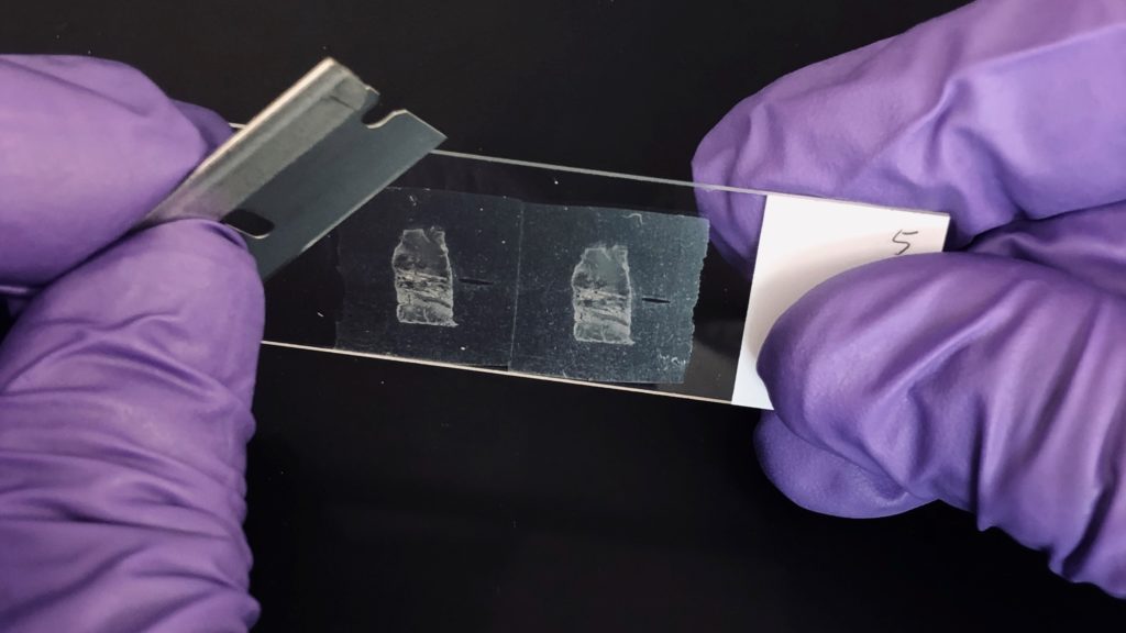
Reliable molecular research starts with reliable sample preparation. Two recently published cancer biology studies illustrate this well, and both studies relied on the Maxwell® RSC platform to extract RNA from formalin-fixed, paraffin-embedded (FFPE) tissue, the archival format that makes up the bulk of clinical pathology material.
Mapping Molecular Targets in a Rare Thyroid Cancer
A 2025 study published in Endocrine Pathology focused on poorly differentiated thyroid carcinoma (PDTC), a rare and aggressive thyroid cancer subtype with limited treatment options once surgery is no longer curative (1). The research question was straightforward but clinically urgent: how many PDTC cases harbor mutations that could be targeted with existing or emerging therapies?
Kierkegaard observed that one of humanity’s enduring tensions is that while life can only be understood backwards, it must be lived forwards. It’s a truth medicine knows intimately: in the treatment that worked until it didn’t, the resistance that arrived without warning, the moment a doctor has to tell a patient that the drug that was helping has stopped. Not because anyone made a mistake, but because the critical knowledge that would have mattered arrived too late, if at all.
A recent paper from the National Cancer Institute is, in a small but meaningful way, science’s pursuit of that elusive foresight: an understanding that emerges early enough, for once, to change what happens next.
The Elegant Idea
For decades, chemotherapy has worked by brute force, flooding the body with toxins designed to kill rapidly dividing cells. The problem is that rapid division isn’t unique to cancer. Hair follicle cells, gut lining cells and immune cells also divide rapidly, which is why patients lose hair, lose energy and become susceptible to infection. Chemotherapy targets a behavior, but the drug has no way to tell a healthy cell from a cancerous one.
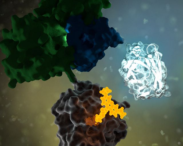
Antibody-drug conjugates (ADCs) change that. Instead of targeting what cancer cells do, they target what cancer cells are. Cancer cells tend to display certain proteins on their surface in far greater numbers than healthy cells do. The antibody is engineered to seek out those proteins specifically. It navigates to its target, binds and waits for the cell to do what cells routinely do: pull it inside. Once there, the cell’s own digestive machinery (the lysosome) breaks down the chemical tether holding the toxin to the antibody, releasing the toxin to kill the cell from within. More than a dozen ADCs have received FDA approval in recent years, and the field is evolving fast.
What the Cell Does Next
But cancer cells don’t simply accept their fate. Even when an ADC delivers its payload perfectly—the antibody finds its target, the cell pulls it inside, the lysosome cuts the tether—a pump embedded in the cell membrane can grab the released toxin and throw it back out before it causes damage.
The delivery worked. The package got ejected anyway.
These pumps—ATP-binding cassette transporters, or more plainly, efflux pumps—are a normal feature of cell biology. Their job is cellular housekeeping, clearing out unwanted or toxic substances before they cause damage. Under the pressure of drug treatment, cancer cells do what life has always done under pressure: the ones best equipped to survive do. The same mechanism that has shaped living things for billions of years now works against the treatment. Not all cancer cells are identical, and the ones that happen to produce more pumps survive while others don’t, gradually shifting the tumor toward resistance.
The brain is one of the most complex and fascinating parts of biology. Thankfully, it’s also remarkably good at protecting itself. When exposed to a pathogen, an injury or even misfolded proteins, microglia and astrocytes function as the central nervous system’s (CNS) primary immune defenders. They mount an inflammatory response by releasing cytokines and working to contain the damage. Yet this same system can malfunction or not resolve, which manifests as devastating consequences.
Chronic neuroinflammation is now recognized as a shared characteristic across some of the most common and difficult-to-treat neurological conditions. A 2023 review in Signal Transduction and Targeted Therapy highlighted the dualistic nature of neuroinflammation: while acute responses serve a protective role, chronic or dysregulated inflammatory signaling can initiate and accelerate neurodegeneration, identifying these pathways as priority targets for therapeutic intervention (Zhang et al., 2023). A 2025 review in Science reinforced this view, noting that within Multiple Sclerosis, disease-modifying therapies targeting neuroinflammation have seen the most clinical success (Shi & Yong, 2025). This could suggest applications within neurological conditions where the same inflammatory mechanisms are at work.
Understanding how and where these inflammatory signals originate in the CNS is an active area of preclinical research. One cytokine being actively studied is IL-6. IL-6 is produced by several cell types, including astrocytes and microglia in the CNS. As a key mediator of inflammatory responses, it mediates pro-inflammatory effects through its trans-signaling, which occurs via soluble IL-6 receptors. Dysregulation of this mechanism may contribute to the chronic neuroinflammation seen in several neurological conditions. Characterizing how and when IL-6 is secreted from CNS cells is an important step toward understanding the neuroinflammatory processes underlying these disorders.
St. Patrick’s Day means different things depending on where you are in the world. In Ireland, it’s a national holiday steeped in culture and tradition: parades, traditional music sessions and, for many, a pint of Guinness accompanied by a hearty “Sláinte” are all part of the day. Here in the American Midwest, we tend to turn that same spirit into a full spectacle. Green everything as far as the eye can see, including somehow an entire river.
Whatever your version of the holiday looks like, there is a lot of fun science behind it. Here is a look at Midwest St. Patrick’s Day through a lab lens.
The Chicago River: Where Orange Becomes Green
Every St. Patrick’s Day, the Chicago Journeymen Plumbers Local 130 heads out on the river and, in roughly 45 minutes, turns a stretch of the Chicago River a surreal emerald green (7, 11). The twist: the dye goes in orange.
The tradition dates to the early 1960s, rooted in a practical idea. Dye had been used to trace leaks and flow in the city’s waterways and someone realized the same concept could be repurposed into a public spectacle (7,11). The exact formula has been kept secret ever since, described only as environmentally friendly and designed to fade after a few hours (1,7,11).
Last spring, my niece and I made a trip to a home improvement store to put together a Mother’s Day planter for my sister. My niece had a clear vision: my sister’s favorite color is blue, so we were going to buy blue flowers. We walked every aisle of the garden center. We checked the annuals, the perennials, and the hanging baskets then left with purple, red, and a grumpy 7-year-old.
It turns out we were not up against a bad selection. We were up against biology.
The Problem with Blue
Blue is one of the rarest colors in the natural world. The food industry is currently finding that out the hard way. There is a good chance you have eaten something blue today. Maybe it was the frosting on a birthday cake, the coating on some M&M’s® candies, or the sports drink in your refrigerator. That blue almost certainly came from a petroleum-based synthetic dye, and for the first time in decades, the food industry is being asked to find something better.
The FDA banned Red Dye No. 3 in January 2025, and pressure has been building around the remaining synthetic dyes ever since, including Blue No. 1 and Blue No. 2. Major food brands have begun announcing plans to reformulate.
There is just one problem. Blue is genuinely, stubbornly hard to make in nature. It turns out that blue has almost nothing to do with color, and almost everything to do with light.
Drug discovery researchers face a fundamental constraint in their work to develop safe, effective therapeutics: the vast majority of the human proteome remains inaccessible to conventional small molecule approaches. Proteins without defined binding pockets, those lacking known chemical probes, and protein targets that fail to translate from biochemical assays into cellular models have long been considered out of reach of standard drug discovery screening tools. As Dixit et al. describe, developing biochemical or cellular assays for all genome-encoded targets “is not scalable and likely impossible as most proteins have ill-defined or unknown activity” — these are what the authors call “the dark undruggable expanses” of the proteome [1].
That gap is now narrowing. Promega Corporation recently launched the TarSeer™ BRETSA™ Target Engagement System, a live-cell target engagement platform designed to bring previously challenging targets within reach of early-stage drug discovery.
The Problem: A Translation Gap in Early Discovery
Drug discovery teams regularly encounter a frustrating disconnect. A compound may show strong binding activity in a biochemical assay, only to fail when tested in a cellular environment. Without target-specific cellular assays, which generally aren’t available for poorly characterized proteins, researchers face difficult choices when deciding which compounds to advance through the drug development pipeline.
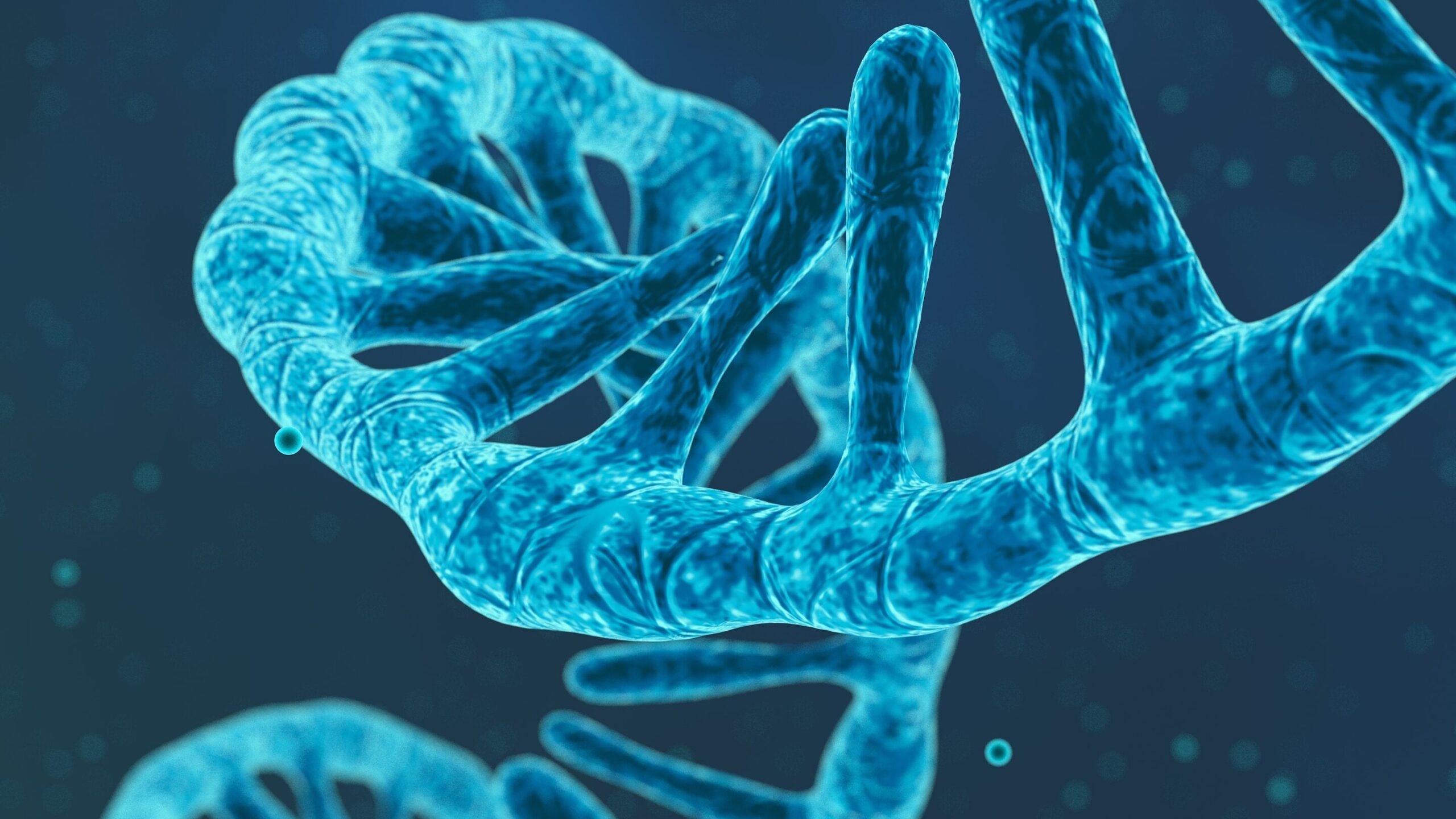
RNA doesn’t just carry genetic instructions—it also interacts with proteins to regulate nearly every aspect of gene expression, from splicing to translation. When those interactions go awry, the consequences can be devastating. In myotonic dystrophy type 1 (DM1), the most common adult-onset muscular dystrophy, a toxic RNA repeat expansion hijacks a critical protein called MBNL1, trapping it in nuclear clumps called foci. This leads to widespread splicing defects and progressive muscle wasting. But studying these toxic interactions inside living cells—and finding small molecules that can disrupt them—has been a significant challenge.
A recent study led by the Scripps Institute may have a solution. The study introduces a NanoBRET™ assay that can monitor the interaction between the expanded CUG RNA repeats and MBNL1 protein in real time, in live cells. Their findings demonstrate how this platform can be used not only to detect disease-driving RNA–protein complexes but also to identify small molecules that break them apart.
Many consider enzymes the workhorses of biochemistry (move over, mitochondria)—catalyzing reactions, breaking down substrates, keeping the machinery of life humming along. But a growing number of researchers are re-envisioning what enzymes can do. Instead of facilitating chemistry, what if enzymes could steer and even guide tiny robots to a tumor?
That’s exactly what’s happening in the rapidly expanding field of enzyme-powered microscopic robots (a.k.a “microrobots”). Microrobots are tiny, engineered devices—often smaller than the width of a human hair—built to perform tasks inside the body that would be difficult or impossible at a larger scale, like delivering drugs to a specific tissue. A recent paper published in Nature Nanotechnology by a team of researchers at California Institute of Technology and the University of Southern California offers a particularly elegant example that we highlight below1.
mRNA-based therapeutics are being explored across a range of applications, including vaccines, protein replacement and immunotherapies (2).
Before any formulation decisions enter the picture, teams need confidence in the RNA itself: that it is the right sequence, right properties and the right purity to behave predictably downstream. That is where it helps to separate drug substance from drug product. The drug substance is the active ingredient intended to deliver a pharmacological effect, while drug product is the finished dosage form that contains that ingredient (6).
This post focuses on what happens upstream, making the mRNA drug substance before formulation. In practical terms, that upstream work spans choosing an mRNA construct, producing it by IVT, and then purifying and analyzing the product so it has the desired quality attributes (5).
When people talk about places where science and technology tend to flourish, a few names surface almost immediately. Silicon Valley, Boston, Seattle, Houston. Cities associated with density, competition and speed.
For many people outside the state, Wisconsin still collapses into a short list of associations: beer, cheese, cold winters, maybe a football team. Biotechnology rarely makes that list.
That hesitation usually has less to do with science itself and more to do with assumptions about where innovation is supposed to live. National Wisconsin Day, celebrated February 15, is a good moment to look past those assumptions and consider what Wisconsin has quietly offered for a long time: an environment and culture that is well-suited for scientific advances.
XWe use cookies and similar technologies to make our website work, run analytics, improve our website, and show you personalized content and advertising. Some of these cookies are essential for our website to work. For others, we won’t set them unless you accept them. To learn more about our approach to Privacy we invite you to Read More
By clicking “Accept All”, you consent to the use of ALL the cookies. However you may visit Cookie Settings to provide a controlled consent.
We use cookies and similar technologies to make our website work, run analytics, improve our website, and show you personalized content and advertising. Some of these cookies are essential for our website to work. For others, we won’t set them unless you accept them. To find out more about cookies and how to manage cookies, read our Cookie Policy.
If you are located in the EEA, the United Kingdom, or Switzerland, you can change your settings at any time by clicking Manage Cookie Consent in the footer of our website.
Necessary cookies are absolutely essential for the website to function properly. These cookies ensure basic functionalities and security features of the website, anonymously.
Cookie
Duration
Description
cookielawinfo-checbox-analytics
11 months
This cookie is set by GDPR Cookie Consent plugin. The cookie is used to store the user consent for the cookies in the category "Analytics".
cookielawinfo-checbox-functional
11 months
The cookie is set by GDPR cookie consent to record the user consent for the cookies in the category "Functional".
cookielawinfo-checbox-others
11 months
This cookie is set by GDPR Cookie Consent plugin. The cookie is used to store the user consent for the cookies in the category "Other.
cookielawinfo-checkbox-advertisement
1 year
The cookie is set by GDPR cookie consent to record the user consent for the cookies in the category "Advertisement".
cookielawinfo-checkbox-necessary
11 months
This cookie is set by GDPR Cookie Consent plugin. The cookies is used to store the user consent for the cookies in the category "Necessary".
cookielawinfo-checkbox-performance
11 months
This cookie is set by GDPR Cookie Consent plugin. The cookie is used to store the user consent for the cookies in the category "Performance".
gdpr_status
6 months 2 days
This cookie is set by the provider Media.net. This cookie is used to check the status whether the user has accepted the cookie consent box. It also helps in not showing the cookie consent box upon re-entry to the website.
lang
This cookie is used to store the language preferences of a user to serve up content in that stored language the next time user visit the website.
viewed_cookie_policy
11 months
The cookie is set by the GDPR Cookie Consent plugin and is used to store whether or not user has consented to the use of cookies. It does not store any personal data.
Analytical cookies are used to understand how visitors interact with the website. These cookies help provide information on metrics the number of visitors, bounce rate, traffic source, etc.
Cookie
Duration
Description
SC_ANALYTICS_GLOBAL_COOKIE
10 years
This cookie is associated with Sitecore content and personalization. This cookie is used to identify the repeat visit from a single user. Sitecore will send a persistent session cookie to the web client.
vuid
2 years
This domain of this cookie is owned by Vimeo. This cookie is used by vimeo to collect tracking information. It sets a unique ID to embed videos to the website.
WMF-Last-Access
1 month 18 hours 24 minutes
This cookie is used to calculate unique devices accessing the website.
_ga
2 years
This cookie is installed by Google Analytics. The cookie is used to calculate visitor, session, campaign data and keep track of site usage for the site's analytics report. The cookies store information anonymously and assign a randomly generated number to identify unique visitors.
_gid
1 day
This cookie is installed by Google Analytics. The cookie is used to store information of how visitors use a website and helps in creating an analytics report of how the website is doing. The data collected including the number visitors, the source where they have come from, and the pages visted in an anonymous form.
Advertisement cookies are used to provide visitors with relevant ads and marketing campaigns. These cookies track visitors across websites and collect information to provide customized ads.
Cookie
Duration
Description
IDE
1 year 24 days
Used by Google DoubleClick and stores information about how the user uses the website and any other advertisement before visiting the website. This is used to present users with ads that are relevant to them according to the user profile.
test_cookie
15 minutes
This cookie is set by doubleclick.net. The purpose of the cookie is to determine if the user's browser supports cookies.
VISITOR_INFO1_LIVE
5 months 27 days
This cookie is set by Youtube. Used to track the information of the embedded YouTube videos on a website.
Performance cookies are used to understand and analyze the key performance indexes of the website which helps in delivering a better user experience for the visitors.
Cookie
Duration
Description
YSC
session
This cookies is set by Youtube and is used to track the views of embedded videos.
_gat_UA-62336821-1
1 minute
This is a pattern type cookie set by Google Analytics, where the pattern element on the name contains the unique identity number of the account or website it relates to. It appears to be a variation of the _gat cookie which is used to limit the amount of data recorded by Google on high traffic volume websites.