G-protein-coupled receptors (GPCRs) are among the most important drug targets in human biology, mediating signals across nearly every physiological system. But not all GPCRs are equally easy to study—especially those that interact with peptide ligands. These ligands tend to be flexible, fast-moving, and hard to trace in live cells by standard methods. Historically, radioligand binding assays have filled this gap, offering a way to measure peptide–receptor interactions with high sensitivity. However, these assays are typically performed using isolated membrane preparations or cells under non-physiological conditions, and they don’t allow for real-time or kinetic measurements.
A recent study published in ACS Chemical Biology by Zhu et. al.1 highlights a modern alternative. Using a HiBiT-tagged version of the galanin receptor (GALR1) and a NanoBRET® assay setup, the researchers were able to monitor peptide binding directly in live cells—without the need for radioactive tracers or cell disruption. GALR1, a neuropeptide receptor involved in feeding, pain, and mood regulation, has historically been difficult to study using traditional tools but has the potential to be impactful in pharmacology. In this blog, we take a closer look at how these researchers applied bioluminescent assays and live-cell imaging to study GPCR-ligand interactions in real time.
A New Approach to Measuring Ligand Binding in Live Cells
Radioligand binding assays offer high sensitivity but come with notable drawbacks: they require specialized facilities to handle radioactive materials, generate hazardous waste, and involve membrane preparations rather than intact cells. This means they can’t capture live-cell dynamics or receptor interactions in their native cellular environment. Radioligand assays are also less applicable to kinetic measurements and high-throughput screening, particularly for peptide ligands with rapid binding kinetics.
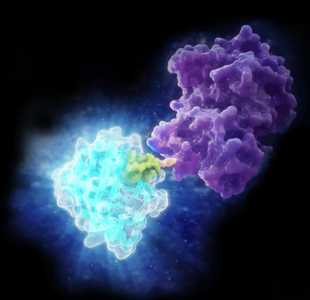
To overcome these limitations, Zhu et al. applied a HiBiT-based NanoBRET® ligand binding assay to study galanin receptor 1 (GALR1) interactions in live cells. HiBiT is an 11–amino acid peptide that produces a bright bioluminescent signal when complemented by LgBiT, a larger subunit derived from NanoLuc® luciferase (learn more about HiBiT technology here). Together with Nano-Glo® substrate, this complementation system generates a luminescent donor signal at the cell surface. When a fluorescently labeled galanin peptide binds the receptor, energy is transferred to the acceptor dye through bioluminescence resonance energy transfer (BRET), producing a signal that reflects receptor occupancy in real time. This cell-based assay enables sensitive, non-radioactive detection of ligand binding in its native environment—without the need for membrane preps, radioactive materials, or wash steps.
From Tracer Design to Biological Validation
To adapt the assay for GALR1 interactions, the authors synthesized six peptide tracers labeled with BODIPY (boron-dipyrromethene), a small, photostable fluorophore well-suited for energy transfer applications like NanoBRET® detection assays. In this study, the dye was conjugated to truncated galanin peptides via short or extended linkers to explore how distance and geometry affect BRET signals.
The six tracers were designed using different peptide backbones: Gal(1–15), M40, and full-length galanin. Each tracer was modified with BODIPY at specific positions. These tracers were evaluated for specificity, binding affinity, and kinetic properties using saturation binding and competitive displacement assays. All six demonstrated the ability to bind GALR1 in live cells, with differences in peptide length and linker design influencing both BRET signal strength and receptor engagement.
Notably, the researchers found that how tightly a tracer bound to the receptor (its binding affinity) closely matched how strongly it activated the receptor. Specifically, peptide tracers that bound more effectively to GALR1 also triggered stronger recruitment of β-arrestin, a key protein involved in GPCR signaling, and greater internalization of the receptor from the cell surface. This suggests that the NanoBRET® assay isn’t just detecting binding but is also predictive of downstream receptor activity. The team confirmed this relationship across three different methods: NanoBRET® to monitor binding, β-arrestin recruitment as a measure of signaling activation, and a HiBiT-based internalization assay to track receptor trafficking.
Bringing Visualization into Focus with GloMax® Galaxy Imager
To confirm that the tracers were binding specifically to GALR1 and that the binding is reversible, the researchers turned to bioluminescent imaging. Using the GloMax® Galaxy Bioluminescence Imager, they visualized NanoBRET® signals in cell lines expressing HiBiT-tagged GALR1. When the fluorescent tracer bound the receptor, energy transfer from the luminescent donor (HiBiT/LgBiT) to the BODIPY-labeled tracer generated a distinct signal—clearly visible in the acceptor channel. This signal diminished when excess unlabeled galanin was added, confirming that the tracer binding was specific and could be competitively displaced.
This reversibility is especially important for ligand-binding assays intended to model real pharmacology, such as competitive displacement screens or kinetic studies. It ensures that the NanoBRET® signal is not only specific to the intended target, but also responsive to biologically meaningful changes—like the presence of a competing ligand. It also supports the utility of this assay for future drug screening efforts, where tracer displacement by candidate compounds would reflect real-time engagement with the receptor in live cells.
This ligand binding assay paired with the images acquired on the GloMax® Galaxy Imager offers this level of resolution without requiring protein overexpression or cell fixation. Unlike traditional fluorescence microscopy, the approach uses native receptor expression and live-cell compatible detection reagents, enabling a clearer, more physiologically relevant view of receptor-ligand interactions. The short, streamlined protocol (no wash steps, no permeabilization) makes this workflow well-suited for both qualitative imaging and quantitative analysis of binding events in real time.
A Step Forward for GPCR Research
Researchers in Zhu et al. demonstrated that HiBiT-based NanoBRET® ligand binding assays can serve as a modern, cell-based alternative to traditional radioligand methods, offering greater specificity, streamlined workflows, and more physiologically-relevant data. By pairing these assays with live-cell imaging on the GloMax® Galaxy Imager, the researchers could measure and visualize ligand binding in real time at the single-cell level.
This novel approach allows researchers to observe and monitor GPCR-ligand interactions with minimal perturbation to the cell. As more labs move toward live-cell, luminescence-based workflows, the combination of NanoBRET® assays and HiBiT technology continues to expand the experimental toolkit in receptor pharmacology and ligand biology.
References
- Zhu, H. et. al. “Development of a HiBiT Peptide-Based NanoBRET Ligand Binding Assay for Galanin Receptor 1 in Live Cells.” ACS Chem. Biol. 2025, 20, 1594–1608. https://doi.org/10.1021/acschembio.5c00166 ↩︎
Anna Bennett
Latest posts by Anna Bennett (see all)
- How Enzymes Are Powering A New Generation of Micro-Robots - February 24, 2026
- A New View of Protein Degradation with HiBiT and Live Cell Imaging - September 23, 2025
- HiBiT-Based NanoBRET® Assay Sheds Light on GPCR–Ligand Binding in Live Cells - July 31, 2025