Photography. From the time the first image was captured almost 200 years ago, people have been using photography techniques to record, improve and expand their world. Joseph Nicéphore Niépce is credited with capturing the very first photograph—a view of outside his window—using a technique called heliography. From that blurry, one-of-a-kind print to the stunning images captured today we can capture using our mobile phones, the advances in personal photography have been phenomenal. But progress has not been limited to capturing images of the world we see, scientists have leveraged these advances into new tools to capture images of the world we can’t see.
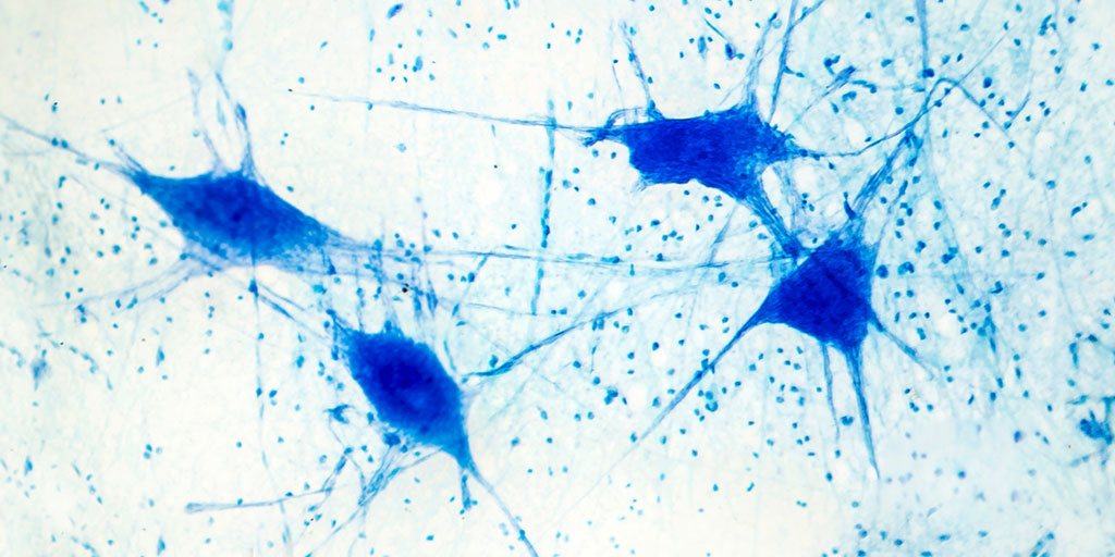
Microcinematography Revealed the Microscopic World
In the early 1900’s, Jean Camandon took a camera developed for cinema films, bolted it to an ultramicroscope and recorded the first video of a microscopic organism, the bacteria Treponema pallidium. Using this technique, the French syphilis researcher was able to demonstrate that the movement of the syphilis disease-causing form of the bacteria was unique from that of the non-disease-causing form. Scientists quickly realized that by using time-lapse microcinematography and then playing the recorded image back at a higher speed they could accelerate the movements. This made slow changes suddenly visible and expanded our understanding of the microscopic world.
In 1943, Dr. Kurt Michel combined time-lapse microcinematography with the newly invented phase-contrast microscope to capture the process of meiosis in spermatocytes of Psophus stridulus (locust). Most of us have probably seen this video at some point in our science education. It remains one of the clearest movies of chromosome behavior during meiosis in live cells.
Digital Imaging Takes Us Inside the Cell
The advent of the self-contained digital camera, which replaced the physical medium of photosensitive film with an electronic photosensor that captured images as identically sized pixels, shepherd in a new era in cell imaging. From this digital technology came widefield microscopy, laser microscopy, bioluminescent and fluorescent imaging. With these techniques, scientist are now able to do anything from localize a protein using whole-body imaging to track the movement of a protein inside a cell.
From the World We See to the World We Don’t
When Niépce captured that first image of the view outside his window, he could never have imagined that less than two hundred years later we would be recording the movements of proteins within a single cell. At its most basic level, photography has been a method of recording an image we see using the action of light or related radiation on a light-sensitive material or photosensor. Beyond that, it has become a valuable tool that is helping us unlock the workings of the world we can’t see.
Additional Reading

Kelly Grooms
Latest posts by Kelly Grooms (see all)
- New Study Suggests Cancer Research Has an Age Problem - April 7, 2026
- Polar Bears, Shrinking Sea Ice and a Scientific Surprise - February 5, 2026
- From Forever Chemicals to Ancient Proteins: Five Science Stories from 2025 - January 8, 2026