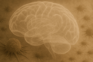
Cancer’s greatest threat is its ability to spread to other tissues—a process known as metastasis. Melanoma, a form of skin cancer, exemplifies this devastating progression. Although treatable when caught early—with surgical removal resulting in over 99% survival at five years—once melanoma metastasizes, five-year survival rates plummet dramatically to around 27%. Even more concerning, melanoma exhibits a particularly high tendency to invade the central nervous system, causing melanoma brain metastases (MBMs) that are incurable and reduce median survival to just 13 months.
To understand metastasis, we need reliable and realistic experimental models. Traditional cell cultures on plastic dishes are limited, failing to replicate the intricate spatial organization and biochemical interactions within living tissues. Animal models are informative but expensive, ethically complex, and not always accurate for human diseases. Addressing this critical gap, Reed-McBain and colleagues (2025) introduced an innovative microphysiological system (MPS) designed to simulate the tumor microenvironment in the brain affected by metastatic melanoma.
In their study, researchers constructed a 3D co-culture platform that integrated dissociated cerebral organoids derived from human induced pluripotent stem cells (iPSCs) with metastatic melanoma cells. This system provided a spatially organized, boundary-free culture, allowing direct interaction between the melanoma cells and neural cells like neurons and astrocytes. After nutrient starvation, viability was measured from each well using an end point ATP-based luminescence assay. With the known location of each well, the researchers were able to generate a luminescence heat-map showing how nutrient stress created a viable rim and necrotic core within the melanoma domain.
Luminescence mapping enabled non-destructive, functional assessment of the microenvironment by quantifying key metabolic indicators such as ATP, glucose, glutamine, and glutamate. This approach allowed for the generation of spatial heatmaps that revealed dynamic changes in viability and metabolism across the platform, offering a scalable solution for high-throughput analysis of tumor-stroma interactions.
Another significant capability illustrated was the detection and visualization of metabolite diffusion, crucial for modeling how tumors disrupt their local environment. Using fluorescent glucose analogs alongside luminescence-based assays, the researchers successfully demonstrated spatial and temporal mapping of glucose diffusion across their MPS. These findings emphasized how metabolite gradients form and change dynamically—insights difficult to glean from traditional cell cultures or endpoint assays.
Perhaps most compellingly, the study showcased the system’s ability to co-culture distinct cell types, capturing interactions crucial to tumor biology. The researchers created melanoma domains within a neuronal scaffold, mirroring tumor-stroma interactions in human brain metastases. They further validated the model by detecting the formation of necrotic cores—areas of cell death due to nutrient deprivation—characteristic of aggressive tumor growth. Additionally, the system captured how melanoma disrupts neural metabolism, notably elevating glutamate levels, which may promote tumor progression and exacerbate neurological damage.
This MPS also accommodated advanced imaging techniques like positron emission tomography (PET), underscoring its potential to bridge in vitro studies and clinical diagnostics. While microscopy offers precise cellular detail, PET imaging complements this by enabling assessment of drug and metabolite diffusion in three-dimensional space, crucial for evaluating therapies.
The study by Reed-McBain et al. highlights how innovative bioengineering can deepen our understanding of pathological and biological processes. This MPS effectively simulates the interactions between tumor and brain cells, offering a promising platform to accelerate therapeutic screening for MBMs. With tools like this, we move closer to unlocking new possibilities in the fight against metastatic melanoma.
Literature Reviewed
Reed-McBain C. et al. (2025) Non-destructive luminescence and PET imaging to monitor tissue microenvironment in microphysiological systems during brain metastasis using dissociated cerebral organoids. Biofabrication 17 035021. DOI:10.1088/1758-5090/ade1fb
Want to measure more metabolites in your research? Explore Promega’s portfolio of bioluminescent assays for metabolic activity, including tools to measure glucose uptake, lactate, glutamine, and more. View the full list of available assays and see if sampling is offered in your region.
Promega Products Used in This Work
The CellTiter-Glo® 2.0 Assay was used to assess cell viability by measuring ATP levels in the MPS model to provide insights into changes in cell health, especially in response to nutrient starvation.
The Glucose-Glo™ Assay was used to measure glucose diffusion within the MPS, enabling the researchers to map glucose gradients spatially and temporally.
The Lactate-Glo™ Assay was used to quantify lactic acid levels providing information about metabolic disruption.
The Glutamine/Glutamate-Glo™ Assay measured glutamine and glutamate, which was indicative of how melanoma cells affected neural metabolism in the MPS.
Michele Arduengo, PhD
Latest posts by Michele Arduengo, PhD (see all)
- What Shelter Dogs Can Tell Us About Emerging Zoonotic Diseases - October 2, 2025
- Developing an Experimental Model System to Understand the Tumor Microenvironment of Melanoma Brain Metastases - September 4, 2025
- IL-6/STAT3-Regulated Long Non-Coding RNA Is Involved in Colorectal Cancer Progression - July 1, 2025