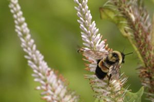
A rusty-patched bumblebee on Culver’s root in the UW–Madison Arboretum. Photo Copyright: SUSAN DAY/UW-MADISON ARBORETUM
Bees have been in the news many times over the past several years. Much of the concern has been focused on the collapse of honey bee colonies because these bees collect nectar to create honey and can be transported for use as pollinators for farmers. Alongside the plight of the honey bee are the declines in the population of native bees in the United States. These bees include insects like the big, fuzzy bumble bees, tiny, iridescent green sweat bees and dark blue mason bees. The native bees live in different conditions. They may be solitary, have a small colony or even nest close together in a communal arrangement, but never in the numbers likely to be seen for a honey bee colony. These lower-density populations can make seeing a change in native bee numbers more difficult. While honey bees have gained the majority of bee decline attention, native bees have suffered dramatic population loss with long-term consequences for the plants they pollinate and the animals that depend upon those plants.
On January 11, 2017, in a landmark decision by the United States Fish and Wildlife Service, the one of the rarest native bees called the rusty-patched bumble bee (Bombus affinis) has been listed as threatened, and this designation will go into effect February 10, 2017. This is the first bee in the U.S. that has been placed on the Endangered Species list. The rusty-patched bumble bee derived its name from the rust-colored patch found on its back.
Studying cellular molecules can be challenging. Some processes are troublesome to study due to the lack of an assay or a complicated assay exists but lacks sensitivity. Membrane proteins in particular are difficult to isolate and characterize. Phosphoglycosyltransferases (PGTs) are transmembrane proteins that transfer phosphosugars onto phospholipids, initiating the synthesis of oligosaccharides in bacterial cell walls. This transfer creates a diphosphate link between a lipid and a sugar and generates UMP as a byproduct. Once this lipid–P–P–sugar linkage occurs, more sugars can be added by glycosyltransferases, generating membrane-based polysaccharides (e.g., peptidoglycan) used for signaling, recognition and defense.
While PGTs have been studied biochemically and an X-ray structure of one member exists, much is still unknown about these enzymes. Overexpressing and purifying membrane proteins remains a challenge, and the conventional PGT assay requires isotope labeled-UDP-sugar donors and is based on the solubility difference between substrate and product to determine enzyme turnover using extraction-based or chromatographic methods. While there are other assays that use fluorescent modified substrates or multienzyme analysis, none of the methods can be applied to all of the diverse PGT enzymes.
All PGTs generate UMP as a byproduct of the transfer of a phosphosugar to a phospholipid. Based on the principle of the luminescent UDP-Glo™ Glycosyltransferase Assay where UDP released during the glycosyltransferase reaction was quantitated, a new luminescent assay called UMP-Glo™ Assay is able to measure the activity of PGT enzymes by adding a single reagent to detect UMP. Das et al. validated this assay by testing PglC, a PGT from Campylobacter jejuni, as well as PglC from Helicobacter pullorum and WecA from Thermatoga maritime and published the results in Scientific Reports. Continue reading “Making It Easier to Investigate PGTs”
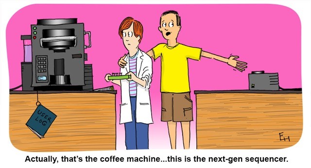
Instruments can make our lives easier in the lab. Place your samples inside an instrument and let it do all the work—isolating nucleic acids or reading and analyzing a multiwell plate—while you walk away to read a new research paper or prepare for the next step in your experiment. However, with the array of machines now available to scientists worldwide, some confusion may result in the laboratory. Has this ever happened to you? Copyright Ed Himelblau
Bubonic plague victims in a mass grave in 18th century France. By S. Tzortzis [Public domain], via Wikimedia CommonsMy last blog post on the Black Death highlighted research that suggested that the reintroduction of Yersinia pestis, the causative agent of the pandemic, originated in Europe during the 14–18th centuries rather than from Asia, the hypothesized origin. In my post, I wrote about my curiosity regarding what an Asian skeleton positive for Y. pestis from that same time period would reveal about the strain or strains that were circulating. Well, a team of researchers has been exploring the issue of strain circulation and an Asian connection, and recently published what they gleaned from additional historic Y. pestis samples in Cell Host & Microbe.
Not every lab has a tried and true transfection protocol that can be used by all lab members. Few researchers will use the same cell type and same construct to generate data. Many times, a scientist may need to transfect different constructs or even different molecules (e.g., short-interfering RNA [siRNA]) into the same cell line, or test a single construct in different cultured cell lines. One construct could be easily transfected into several different cell lines or a transfection protocol may work for several different constructs. However, some cells like primary cells can be difficult to transfect and some nucleic acids will need to be optimized for successful transfection. Here are some tips that may help you improve your transfection success.
Transfect healthy, actively dividing cells at a consistent cell density. Cells should be at a low passage number and 50–80% confluent when transfected. Using the same cell density reduces variability for replicates. Keep cells Mycoplasma-free to ensure optimal growth.
Transfect using high-quality DNA. Transfection-quality DNA is free from protein, RNA and chemical contamination with an A260/A280 ratio of 1.7–1.9. Prepare purified DNA in sterile water or TE buffer at a final concentration of 0.2–1mg/ml.
RNA molecules have become a hot topic of research. While I was taught about messenger RNA (mRNA), ribosomal RNA (rRNA) and transfer RNA (tRNA), many more varieties have come into the nomenclature after I graduated with my science degrees. Even more interesting, these RNAs do not code for a protein, but instead have a role in regulating gene expression. From long non-coding RNA (lncRNA) to short interfering RNA (siRNA), microRNA (miRNA) and small nucleolar RNA (snoRNA), these classes of RNAs affect protein translation, whether by hindering ribosomal binding, targeting mRNA for degradation or even modifying DNA (e.g., methylation). This post will cover the topic of microRNAs, explaining what they are, how researchers understand their function and role in metabolism, cancer and cardiovascular disease, and some of the challenges in miRNA research.
What are microRNAs? MicroRNAs (miRNAs) are short noncoding RNAs 19–25 nucleotides long that play a role in protein expression by regulating translation initiation and degrading mRNA. miRNAs are coded as genes in DNA and transcribed by RNA polymerase as a primary transcript (pri-miRNA) that is hundreds or thousands of nucleotides long. After processing with a double-stranded RNA-specific nuclease, a 70–100 nucleotide hairpin RNA precursor (pre-miRNA) is generated and transported from the nucleus into the cytoplasm. Once in the cytoplasm, the pre-miRNA is cleaved into an 18- to 24-nucleotide duplex by ribonuclease III (Dicer). This cleaved duplex associates with the RNA-induced silencing complex (RISC), and one strand of the miRNA duplex remains with RISC to become the mature miRNA.
Honey bees are hard-working insects. Their pollination services are in such demand, humans tow hundreds of hives carrying millions of bees around in the back of semitrucks to bring honey bees to various locations such as California almond groves. Humans are also quite partial to the bee colony winter energy storage also known as honey. So while honey bees work hard to collect pollen and nectar from blooming plants and trees and store honey for the winter, humans insist on robbing the colony’s store of delicious sweetener for their own uses. Recent reports of high mortality in honey bee colonies has caused concern in many beekeepers who manage European honey bee apiaries for honey production and pollination services. These severe depletion of honey bee colonies have been attributed to the parasitic mite Varroa destructor in the colony, not only feeding off the larvae and pupae brooding in the colony but also transmitting viruses carried by the mite. Bee nutrition is important for the pollinators especially when overwintering in the hive. Without adequate nutrition, a colony may become weak and succumb to parasite or disease pressure, unable to survive until nectar and pollen are available in the spring. A study was recently published in PLOS ONE that examined how the landscape around Midwestern honeybee hives affected the ability of bees to overwinter and assessed their health by measuring levels of Varroa mites and honey bee viruses. Continue reading “How Do Agricultural Landscapes Affect Bee Health?”
Macrophages. By NIAID ) [CC BY 2.0 , via Wikimedia Commons
Many think of glucose as something diabetics have to test each day using a blood monitor, or a quick energy boost for someone exercising intensely. However, the simple sugar glucose, a monosaccharide, fuels most of the cells in our bodies. Disaccharides that contain glucose (e.g., sucrose is comprised of glucose and fructose) and glucose polymers (e.g., starch and glycogen) are carbohydrates that are consumed by organisms from bacteria to humans to produce energy. These carbohydrates are broken down into component monosaccharides like glucose and lactose. The process of glycolysis generates the energy currency of cells, ATP, as well as precursor molecules for nucleotides, lipids and amino acids. Because glucose is the cell fuel source, the uptake of glucose and its subsequent metabolism is increased by cells that divide rapidly like cancer cells. The more energy and precursor molecules the cancer cell can create for itself, the more rapidly the tumor can grow.
Because glucose metabolism is central to cellular functioning, changes that decrease glucose uptake or increase glycolysis have a widespread effect on on both the cells and organism. How does a simple sugar molecule create such broad effects on health? For example, diabetes results from the inability to store glucose because of a lack of insulin, a hormone that draws glucose from the blood and stores it as glycogen in the liver, muscles and adipose tissue. High levels of sugar in the blood negatively affect the body over the long term, damaging blood vessels and eyesight, making the kidneys work harder to excrete the excess sugar and increasing the risk of stroke and coronary artery disease. Because cancer cells have such a high metabolic demand for glucose, many of the mutations in cancers affect pathways that regulate glucose uptake and glucose breakdown, allowing the cancer cells to survive and grow, crowding out nearby normal cells.
Glucose metabolism is altered by processes other than mutations or an reduced production of a hormone. Throughout its life cycle, a cell will vary its requirements for glucose. For example, the cells that comprise our innate immune response are typically in a quiescent or steady state. However, when these immune cells encounter an foreign invader, they become activated and increase their demand for glucose. To respond to a potential pathogen, the activated cells need glucose to fuel cell proliferation and the production of cytokines, chemicals that activate other immune cells and initiate an inflammatory response. The typical signs of inflammation are red inflamed area that may be painful to the touch, such as a cut that becomes infected. Most inflammation resolves when the infection is eliminated, leaving behind whole skin in the instance of a cut, and the activated immune cells become quiescent again.
An Interesting Observation about Glucose Metabolism in M2 Macrophages
Glucose uptake, immunity and metabolism are cellular pathways that are intertwined such that understanding how glucose is utilized in macrophages illuminates gene induction and regulation in activated macrophages. In a recently published eLife article, Covarrubias et al. studied how activation of murine bone marrow-derived macrophages (BMDMs) by interleukin-4 (IL-4), a signaling cytokine, altered glucose metabolism in the cells and regulated a subset of genes involved in macrophage activation.
Yersinia pestis. See page for author [Public domain], via Wikimedia CommonsWhile scientists using ancient DNA analysis are learning how Yersinia pestis developed over time into the causative agent of three worldwide pandemics, there is still much to learn about the early hours and days of an organism infected with the plague. Y. pestis still infects humans so any insight into disease progression is useful for determining treatment timing and even developing novel treatments to supplement or replace antibiotics. A 2012 study observed how Y. pestis injected into mice spread throughout the body using bioluminescent imaging to track the infection. More recent research reported in PLOS ONE used a more real-world route of infection by introducing an aerosolized Y. pestis to a nonhuman primate model and tracking the transcripts altered during the first 42 hours of infection. Continue reading “Analyzing the Effects of Yersinia pestis Infection on Gene Expression”
Occasionally, time in the lab passes slowly. There is a two-hour incubation and nothing can be done until the timer goes off. Our science cartoonist Ed Himelblau has illustrated what some creative lab members may have done to fill this time, but is not advised to do:
Copyright Ed Himelblau
To see additional lab shenanigans, peruse the collection of humorous cartoons in our Cartoon Lab.
XWe use cookies and similar technologies to make our website work, run analytics, improve our website, and show you personalized content and advertising. Some of these cookies are essential for our website to work. For others, we won’t set them unless you accept them. To learn more about our approach to Privacy we invite you to Read More
By clicking “Accept All”, you consent to the use of ALL the cookies. However you may visit Cookie Settings to provide a controlled consent.
We use cookies and similar technologies to make our website work, run analytics, improve our website, and show you personalized content and advertising. Some of these cookies are essential for our website to work. For others, we won’t set them unless you accept them. To find out more about cookies and how to manage cookies, read our Cookie Policy.
If you are located in the EEA, the United Kingdom, or Switzerland, you can change your settings at any time by clicking Manage Cookie Consent in the footer of our website.
Necessary cookies are absolutely essential for the website to function properly. These cookies ensure basic functionalities and security features of the website, anonymously.
Cookie
Duration
Description
cookielawinfo-checbox-analytics
11 months
This cookie is set by GDPR Cookie Consent plugin. The cookie is used to store the user consent for the cookies in the category "Analytics".
cookielawinfo-checbox-functional
11 months
The cookie is set by GDPR cookie consent to record the user consent for the cookies in the category "Functional".
cookielawinfo-checbox-others
11 months
This cookie is set by GDPR Cookie Consent plugin. The cookie is used to store the user consent for the cookies in the category "Other.
cookielawinfo-checkbox-advertisement
1 year
The cookie is set by GDPR cookie consent to record the user consent for the cookies in the category "Advertisement".
cookielawinfo-checkbox-necessary
11 months
This cookie is set by GDPR Cookie Consent plugin. The cookies is used to store the user consent for the cookies in the category "Necessary".
cookielawinfo-checkbox-performance
11 months
This cookie is set by GDPR Cookie Consent plugin. The cookie is used to store the user consent for the cookies in the category "Performance".
gdpr_status
6 months 2 days
This cookie is set by the provider Media.net. This cookie is used to check the status whether the user has accepted the cookie consent box. It also helps in not showing the cookie consent box upon re-entry to the website.
lang
This cookie is used to store the language preferences of a user to serve up content in that stored language the next time user visit the website.
viewed_cookie_policy
11 months
The cookie is set by the GDPR Cookie Consent plugin and is used to store whether or not user has consented to the use of cookies. It does not store any personal data.
Analytical cookies are used to understand how visitors interact with the website. These cookies help provide information on metrics the number of visitors, bounce rate, traffic source, etc.
Cookie
Duration
Description
SC_ANALYTICS_GLOBAL_COOKIE
10 years
This cookie is associated with Sitecore content and personalization. This cookie is used to identify the repeat visit from a single user. Sitecore will send a persistent session cookie to the web client.
vuid
2 years
This domain of this cookie is owned by Vimeo. This cookie is used by vimeo to collect tracking information. It sets a unique ID to embed videos to the website.
WMF-Last-Access
1 month 18 hours 24 minutes
This cookie is used to calculate unique devices accessing the website.
_ga
2 years
This cookie is installed by Google Analytics. The cookie is used to calculate visitor, session, campaign data and keep track of site usage for the site's analytics report. The cookies store information anonymously and assign a randomly generated number to identify unique visitors.
_gid
1 day
This cookie is installed by Google Analytics. The cookie is used to store information of how visitors use a website and helps in creating an analytics report of how the website is doing. The data collected including the number visitors, the source where they have come from, and the pages visted in an anonymous form.
Advertisement cookies are used to provide visitors with relevant ads and marketing campaigns. These cookies track visitors across websites and collect information to provide customized ads.
Cookie
Duration
Description
IDE
1 year 24 days
Used by Google DoubleClick and stores information about how the user uses the website and any other advertisement before visiting the website. This is used to present users with ads that are relevant to them according to the user profile.
test_cookie
15 minutes
This cookie is set by doubleclick.net. The purpose of the cookie is to determine if the user's browser supports cookies.
VISITOR_INFO1_LIVE
5 months 27 days
This cookie is set by Youtube. Used to track the information of the embedded YouTube videos on a website.
Performance cookies are used to understand and analyze the key performance indexes of the website which helps in delivering a better user experience for the visitors.
Cookie
Duration
Description
YSC
session
This cookies is set by Youtube and is used to track the views of embedded videos.
_gat_UA-62336821-1
1 minute
This is a pattern type cookie set by Google Analytics, where the pattern element on the name contains the unique identity number of the account or website it relates to. It appears to be a variation of the _gat cookie which is used to limit the amount of data recorded by Google on high traffic volume websites.
![Bubonic plague victims in a mass grave in 18th century France. By S. Tzortzis [Public domain], via Wikimedia Commons](https://commons.wikimedia.org/wiki/File%3ABubonic_plague_victims-mass_grave_in_Martigues%2C_France_1720-1721.jpg)


 Honey bees are hard-working insects. Their pollination services are in such demand, humans tow hundreds of hives carrying millions of bees around in the back of semitrucks to bring honey bees to various locations such as California almond groves. Humans are also quite partial to the bee colony winter energy storage also known as honey. So while honey bees work hard to collect pollen and nectar from blooming plants and trees and store honey for the winter, humans insist on robbing the colony’s store of delicious sweetener for their own uses. Recent reports of high mortality in honey bee colonies has caused concern in many beekeepers who manage European honey bee apiaries for honey production and pollination services. These severe depletion of honey bee colonies have been attributed to the parasitic mite Varroa destructor in the colony, not only feeding off the larvae and pupae brooding in the colony but also transmitting viruses carried by the mite. Bee nutrition is important for the pollinators especially when overwintering in the hive. Without adequate nutrition, a colony may become weak and succumb to parasite or disease pressure, unable to survive until nectar and pollen are available in the spring. A study was recently published in PLOS ONE that examined how the landscape around Midwestern honeybee hives affected the ability of bees to overwinter and assessed their health by measuring levels of Varroa mites and honey bee viruses.