What can you do with a small, super bright luciferase? Amazing things. We’ve highlighted many of the papers and new applications that NanoLuc® luciferase has enabled on this blog. While NanoLuc® luciferase was first introduced as a reporter enzyme to assess promoter activity, its capabilities have expanded far beyond a genetic reporter, creating bioluminescent tools used to study endogeneous protein dynamics, target engagement, protein degradation, immunodetection and more. So where did the NanoLuc luciferase come from and how does one enzyme power so many research capabilities? Read further for a primer on the various technologies and applications developed from this enzyme over the last 10 years.
Asian elephants with babies in Planckendael zoo, Muizen near Mechelen, Flanders, Belgium. Image copyright: Ad Meskens [CC BY-SA 4.0 (https://creativecommons.org/licenses/by-sa/4.0)] via Wikimedia Commons
Wildlife conservation is a major focus around the world. With habitat loss and climate change, Asian elephant populations are under severe pressure. Add in an infectious disease that is fatal to the young and you have a recipe for disaster. Even with efforts to breed the endangered Asian elephants in zoos to build the population, elephant endotheliotropic herpesvirus (EEHV) thwarts conservation efforts. EEHV causes hemorrhagic disease in Asian elephants younger than 10 years old, a disease with rapid onset and high mortality. In fact, some numbers indicate EEHV is the cause of death for at least 25% of Asian elephants born in zoos and the wild globally.
You have identified and cloned your protein of interest, but you want to explore its function. A protein fusion tag might help with your investigation. However, choosing a tag for your protein depends on what experiments you are planning. Do you want to purify the protein? Would you like to identify interacting proteins by performing pull-down assays? Are you interested in examining the endogenous biology of the protein? Here we cover the advantages and disadvantages of some protein tags to help you select the one that best suits your needs.
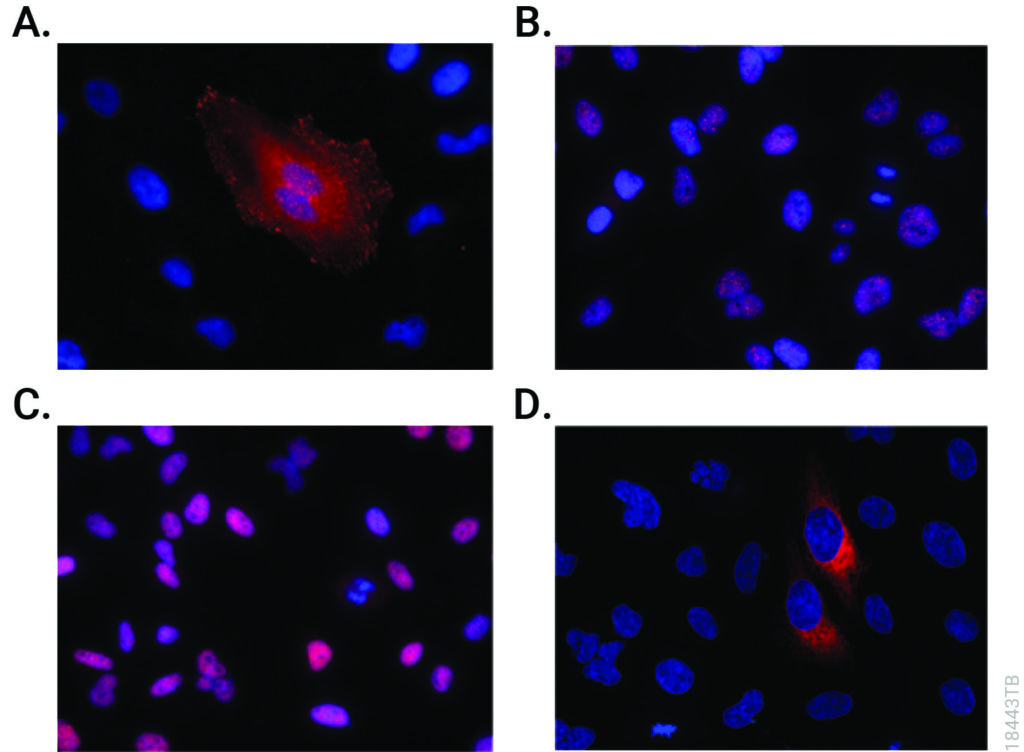
CRISPR-Cas9 editing knocked-in HiBiT at the endogenous locus of proteins with varying subcellular localization. Fixed CRISPR-modified clones or pools of cells were imaged by immunofluorescent staining using the Anti-HiBiT Monoclonal Antibody (red) and Hoechst dye (blue). Panel A. VCL-HiBiT pool. Panel B. SMARCA4-HiBiT clone. Panel C. HDAC2-HiBiT clone. Panel D. HSP90B1-HiBiT pool.
Affinity Tags
The most commonly used protein tags fall under the category of affinity tags. This means that the tag binds to another molecule or metal ion, making it easy to purify or pull down your protein of interest. In all cases, the tag will be fused to your protein of interest at either the amino (N) or carboxy (C) terminus by cloning into an expression vector. This protein fusion can then be expressed in cells or cell-free systems, depending on the promoter the vector contains.
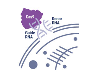
Ribonucleoprotein complex with Cas9, guide RNA and donor ssDNA. Copyright Promega Corporation.
With the advent of genome editing using CRISPR-Cas9, researchers have been excited by the possibilities of precisely placed edits in cellular DNA. Any double-stranded break in DNA, like that induced by CRISPR-Cas9, is repaired by one of two pathways: Non-homologous end joining (NHEJ) or homology-directed repair (HDR). Using the NHEJ pathway results in short insertions or deletions (indels) at the break site, so the HDR pathway is preferred. However, the low efficiency of HDR recombination to insert exogenous sequences into the genome hampers its use. There have been many attempts at boosting HDR frequency, but the methods compromise cell growth and behave differently when used with various cell types and gene targets. The strategy employed by the authors of an article in Communications Biology tethered the DNA donor template to Cas9 complexed with the ribonucleoprotein and guide RNA, increasing the local concentration of the donor template at the break site and enhancing homology-directed repair. Continue reading “All You Need is a Tether: Improving Repair Efficiency for CRISPR-Cas9 Gene Editing”
Yersinia pestis. U.S. Center for Disease Control [Public domain], via Wikimedia Commons.Human teeth play a key role in our understanding of how organisms evolve. Whenever a possible new member of the hominid family is uncovered, the shape and number of teeth are used to place that individual in the family tree. Teeth also harbor information about pathogens that have plagued humans for millennia. Because bacteria use our bloodstream as a transport system, protected places that can preserve DNA—like the pulp of teeth—are a rich medium for uncovering information about humans and the microbes that infected them.
Teeth have been the choice for identifying the infectious agent behind the Plague of Justinian in the sixth century and the Black Plague in the 14th century. In fact, Yersinia pestis, the bacterium responsible for these plagues, has infected humans as far back as the Neolithic. But what can we learn about the pandemic strain or strains of Y. pestis described in historical records? A team of researchers from Europe and the US, many of whom have been delving into the history of Y. pestis for the last decade, wanted to further investigate the Plague of Justinian. They studied bacterial DNA extracted from human remains found in Western European communal graves that were dated to around 541–750, the period of the historically documented Plague of Justinian. Their investigation examined the bacteria’s diversity and how far it spread during this “First Pandemic” of plague. Continue reading “Delving into the Diversity of The Plague of Justinian”
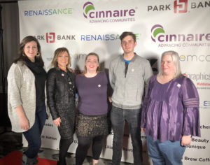
The five-member team at the Read(y) To Wear event.
What do fashion, paperboard product packaging and literacy have in common? Answer: The Read(y) to Wear submission from a team of Promega employees for an event put on by the Madison Reading Project. With a challenge that stated teams need to make a garment mostly of paper, the resulting creations would be displayed on a runway as part of a charitable evening for an organization dedicated to bringing books to children.
Volunteering to be part of what became a five-person team to create a wearable garment from paper was the easy part. Our first few meetings we were experimenting with ideas and techniques using paper we could access on campus: Print catalogs, discarded books and our prototype product kit boxes. It was the kit boxes with the David Goodsell imagery that inspired our ideas to create a suit of armor. The paperboard boxes protect the products we ship to customers like a suit of armor protects warriors in battle. Continue reading “Cardboard Couture: From Conception to Runway Debut”
Yersinia pestis. See page for author [Public domain], via Wikimedia Commons
In recent years, scientists have been able to refine their molecular tools to resurrect ancient DNA from human graves and determine that yes, Yersinia pestis was the causative agent for the Black Death in the 14th century and the Plague of Justinian in the 6th century. As more and more human graves have been uncovered, their DNA has revealed many secrets that scientists even ten years ago were unable to discover. With the ability to sequence entire genomes of bacteria that died with their hosts hundreds and even thousands of years ago, researchers are exploring the rise and possible spread of Y. pestis. Each new member sequence adds to the Y. pestis family tree, pinpointing the origin of this bacteria as it diverged from its ancestor Y. pseudotuberculosis. Peering into the past, scientists have been able to track down a strain of Y. pestis from individuals in a Swedish passage grave that is basal to known strains and that the authors of a Cell article suggest has interesting implications.
This pathogenic journey into history started by analyzing ancient DNA data sets from the teeth of individuals present in a communal passage grave in Gökhem parish, located in western Sweden, for any disease-causing microbial sequences that might be present. Y. pestis was flagged in one 20-year-old female dated 4,867–5,040 years ago. The bacterial sequences from this individual, named Gok2, were more closely aligned with Y. pestis than the Y. pseudotuberculosis reference genome.
Glucose is an energy metabolite necessary for cellular survival and growth whether or not the cell is part of a tumor. Not only do cancer cells switch from oxidative phosphorylation to aerobic glycolysis (the Warburg effect) to gain more glucose, a hallmark of cancer, but they also increase the amount of glucose taken up from the surrounding extracellular space. However, the lack of glucose can have a negative effect on cells, causing them to become apoptotic in the absence of this metabolite. Cancer cells have methods to get around the requirement for glucose, including upregulating glucose transporters to improve access to the energy metabolite. In this Redox Biology article, researchers describe how activating androgen receptor in response to a lack of glucose affects the amount of GLUT1 expressed on prostate cancer cells, making the cells resistant to glucose deprivation.
To set the stage, two prostate cancer cell lines, LNCaP, an androgen-sensitive cell line, and LNCaP-R, an androgen-insensitive cell line, were deprived of glucose. Both cell lines showed signs of cell death, but LNCaP-R cells died in greater numbers. To probe how LNCaP cells died, several inhibitors (a pan-caspase inhibitor, two necroptosis inhibitors and a ferroptosis inhibitor) were added but did not change the way the cells died. However, an autophagy inhibitor enhanced cell death, suggesting the cells were necrotic not apoptotic. Teasing apart if the necrosis of LNCaP cells was due to glucose availability or merely disrupted glycolysis, the glucose analog 2DG was added to the medium with glucose. The cells survived when treated with 2DG, suggesting it was the absence of glucose that induced necrosis. When LNCaP cells were cultivated in medium that replaced glucose with mannose or fructose, the cells survived, another point in favor of sugar depletion causing cell death.
“The Coronation of the Virgin” tempera on panel by Fra Angelico [Public domain], via Wikimedia Commons
Chicken eggs are widely found in most grocery stores. They are a cheap and unassuming source of protein, easy to cook as you desire (e.g., fried, scrambled, hard boiled) or use as a binder for other food items (e.g., meatloaf, cakes, cookies). One reason I keep laying hens myself is not only for fresh eggs but also to have egg shells other colors than white, the predominant color sold in US grocery stores. However, did you know that these humble eggs have uses that don’t end up in the stomach and instead, are a feast for the eyes? I was introduced to the concept of egg tempera, a medium used for painting, by my colleague, Karen Stakun, artist and manager of our graphics department during a discussion about chickens. I was intrigued by the concept.
Pale purple asters and milkweed. Copyright S. Klink.
Surrounding my mowed lawn is a wild, mostly uncultivated space that currently has goldenrod blooming with tall asters starting to blossom. Every day when I pass these flowers, I see bumblebees, butterflies and other insects collecting the nectar to eat or store for the winter. Last year, when a section of soil was disturbed during construction of a building, I decided to seed the area with native wildflowers rather than grass. (I am not a fan of mowing the lawn.) Watching the series of flowers bloom over the late spring to autumn has been beautiful, colorful and full of tiny moments of joy. Not only do I see insects enjoying the flowering plants, but birds will land on the taller greenery, sometimes just resting, sometimes collecting seeds. I am not sure who has been startled more often, me or the birds when I walk by, flushing a bird from the thicket of tall plants.
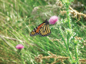
Monarch butterfly on thistle photographed in the prairie at Promega headquarters in Madison, WI. Copyright Promega Corporation.
Where some people might see wild, unruly areas, I see Monarch butterflies on their daily flight, fluttering above me and the “weeds”. I have even been lucky enough to find Monarch caterpillars munching on milkweed, a common plant in my wild space. Despite my efforts, I have a lot of tall ragweed appearing in my yard, but have discovered that birds love the seeds, including my chickens, and squirrels will remove and eat the leaves. In addition, I see fireflies in early June through late August, many I find hanging out on the shady greenery during the day before their light display at night. Continue reading “In Defense of Wild Spaces in the Yard”
XWe use cookies and similar technologies to make our website work, run analytics, improve our website, and show you personalized content and advertising. Some of these cookies are essential for our website to work. For others, we won’t set them unless you accept them. To learn more about our approach to Privacy we invite you to Read More
By clicking “Accept All”, you consent to the use of ALL the cookies. However you may visit Cookie Settings to provide a controlled consent.
We use cookies and similar technologies to make our website work, run analytics, improve our website, and show you personalized content and advertising. Some of these cookies are essential for our website to work. For others, we won’t set them unless you accept them. To find out more about cookies and how to manage cookies, read our Cookie Policy.
If you are located in the EEA, the United Kingdom, or Switzerland, you can change your settings at any time by clicking Manage Cookie Consent in the footer of our website.
Necessary cookies are absolutely essential for the website to function properly. These cookies ensure basic functionalities and security features of the website, anonymously.
Cookie
Duration
Description
cookielawinfo-checbox-analytics
11 months
This cookie is set by GDPR Cookie Consent plugin. The cookie is used to store the user consent for the cookies in the category "Analytics".
cookielawinfo-checbox-functional
11 months
The cookie is set by GDPR cookie consent to record the user consent for the cookies in the category "Functional".
cookielawinfo-checbox-others
11 months
This cookie is set by GDPR Cookie Consent plugin. The cookie is used to store the user consent for the cookies in the category "Other.
cookielawinfo-checkbox-advertisement
1 year
The cookie is set by GDPR cookie consent to record the user consent for the cookies in the category "Advertisement".
cookielawinfo-checkbox-necessary
11 months
This cookie is set by GDPR Cookie Consent plugin. The cookies is used to store the user consent for the cookies in the category "Necessary".
cookielawinfo-checkbox-performance
11 months
This cookie is set by GDPR Cookie Consent plugin. The cookie is used to store the user consent for the cookies in the category "Performance".
gdpr_status
6 months 2 days
This cookie is set by the provider Media.net. This cookie is used to check the status whether the user has accepted the cookie consent box. It also helps in not showing the cookie consent box upon re-entry to the website.
lang
This cookie is used to store the language preferences of a user to serve up content in that stored language the next time user visit the website.
viewed_cookie_policy
11 months
The cookie is set by the GDPR Cookie Consent plugin and is used to store whether or not user has consented to the use of cookies. It does not store any personal data.
Analytical cookies are used to understand how visitors interact with the website. These cookies help provide information on metrics the number of visitors, bounce rate, traffic source, etc.
Cookie
Duration
Description
SC_ANALYTICS_GLOBAL_COOKIE
10 years
This cookie is associated with Sitecore content and personalization. This cookie is used to identify the repeat visit from a single user. Sitecore will send a persistent session cookie to the web client.
vuid
2 years
This domain of this cookie is owned by Vimeo. This cookie is used by vimeo to collect tracking information. It sets a unique ID to embed videos to the website.
WMF-Last-Access
1 month 18 hours 24 minutes
This cookie is used to calculate unique devices accessing the website.
_ga
2 years
This cookie is installed by Google Analytics. The cookie is used to calculate visitor, session, campaign data and keep track of site usage for the site's analytics report. The cookies store information anonymously and assign a randomly generated number to identify unique visitors.
_gid
1 day
This cookie is installed by Google Analytics. The cookie is used to store information of how visitors use a website and helps in creating an analytics report of how the website is doing. The data collected including the number visitors, the source where they have come from, and the pages visted in an anonymous form.
Advertisement cookies are used to provide visitors with relevant ads and marketing campaigns. These cookies track visitors across websites and collect information to provide customized ads.
Cookie
Duration
Description
IDE
1 year 24 days
Used by Google DoubleClick and stores information about how the user uses the website and any other advertisement before visiting the website. This is used to present users with ads that are relevant to them according to the user profile.
test_cookie
15 minutes
This cookie is set by doubleclick.net. The purpose of the cookie is to determine if the user's browser supports cookies.
VISITOR_INFO1_LIVE
5 months 27 days
This cookie is set by Youtube. Used to track the information of the embedded YouTube videos on a website.
Performance cookies are used to understand and analyze the key performance indexes of the website which helps in delivering a better user experience for the visitors.
Cookie
Duration
Description
YSC
session
This cookies is set by Youtube and is used to track the views of embedded videos.
_gat_UA-62336821-1
1 minute
This is a pattern type cookie set by Google Analytics, where the pattern element on the name contains the unique identity number of the account or website it relates to. It appears to be a variation of the _gat cookie which is used to limit the amount of data recorded by Google on high traffic volume websites.