Looking Back: Cell-Free Expression Systems Helped to Characterize Proteins Involved in Hypoxia Response

Structur of a HIF-1a-pVHL-ElonginB-ElonginC complex
Structure of a HIF-1a-pVHL-ElonginB-ElonginC complex

William G. Kaelin Jr., Sir Peter J. Ratcliffe and Gregg L. Semenza were awarded the 2019 Nobel Prize in Physiology or Medicine for their discoveries of how cells sense and adapt to oxygen availability.

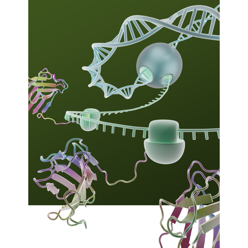
Kaelin and Ratcliffe’s labs focused their efforts on the transcription factor HIF (hypoxia-inducible factor). This transcription factor is critical in the cellular adaptation of to changes in oxygen availability.

When oxygen levels are elevated cells contain very little HIF. Ubiquitin is added to the HIF protein via the VHL complex and it is degraded in the proteasome.  When oxygen levels are low (hypoxia) the amount of HIF increases.

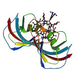
In 2001 both groups published articles characterizing the interaction between VHL and HIF, and these articles were referenced by the Nobel Prize Organization in their press release about this year’s award. (1,2). Both studies demonstrated that under the normal oxygen conditions hydroxylation of proline residue P564 enabled VHL to recognize and bind to HIF.

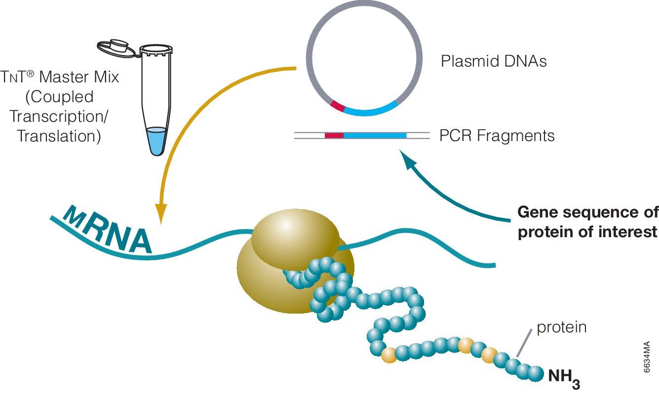
The use of cell free expression (i.e., TNT Coupled Transcription/Translation System) by both labs was key in the characterization of the VHL:HIF interaction The labs utilized HIF and VHL 35-S labeled proteins generated via the TNT system under both normal or in a hypoxic work station to:

  • Determine the affect of ferrous chloride and cobaltous chloride on the interaction
  • Map the specific region of HIF required for the interaction to occur (556-574)
  • Determine the effect of HIF point mutations on the interaction
  • Use synthetic peptides to block the interaction
  • Conclude that a factor in mammalian cells was necessary for the interaction to occur.

Literature Cited

  1. Ivan, M et al. (2001) HIF Targeted for VHL-Mediated Destruction by Proline Hydroxylation: Implications for O2 Sensing. Science 292: 464–67.
  2. Jaakkola, P. et al. (2001) Targeting of HIF-α to the von Hippel-Lindau Ubiquitylation of Complex by O2– Regulated Prolyl Hydroxylation. Science 202, 468–72 .

Related Posts

Characterizing Compound Binding in Cell-Free Systems

Dioxins (e.g., 2,3,7,8-Tetrachlorodibenzo-p-dioxin, TCDD) and related compounds (DRCs) are persistent environmental pollutants that gradually accumulate through the food chain, mainly in the fatty tissues of animals. Dioxins are highly toxic and can cause reproductive and developmental problems, damage the immune system, interfere with hormones and also cause cancer. This broad range of toxic and biological effects of DRCs is mostly mediated by the aryl hydrocarbon receptor (AHR).

In animal cells, DRCs bind to AHR in the cytoplasm and then translocate into the nucleus, where they affect the transcription of multiple target genes, including xenobiotic-metabolizing enzymes, such as CYP1A isozymes. AHR is also involved in immune system maintenance, protein degradation and cell proliferation.

The jungle crow (Corvus macrorhynchos) has been considered a suitable indicator for monitoring environmental chemicals such as DRCs. While mammals only have one AHR form, avian species have multiple AHR isoforms such as AHR1 and AHR2. To unveil the functional diversity of multiple avian AHR isoforms in terms of their contribution to responses to DRCs a recent study by Kim et al. investigated the molecular and functional characteristics of jungle crow AHR isoforms, cAHR1 and jcAHR2 (1).

cAHR1 and jcAHR2 proteins were synthesized using AHR proteins were synthesized using the TnT Quick-Coupled Reticulocyte Lysate System  to examine whether these jcAHRs have the potential to bind to TCDD. TCDD-binding affinity of the in vitro-expressed jcAHR protein was analyzed using the velocity sedimentation assay with a sucrose gradient.

The results demonstrate that both jcAHR1and jcAHR2 are capable of binding to TCDD.

Reference
Kim, E-Y (2019) The aryl hydrocarbon receptor 2 potentially mediates cytochrome P450 1A induction in the jungle crow (Corvus macrorhynchos). Ecotoxicology and Environmental Safety 171. 99–111

Tetanus Neurotoxin: Potential Mechanism for Drug Delivery

Tetanus neurotoxin (TeNT), produced by Clostridium tetani, is one of the most potent neurotoxins in humans. TeNT causes tetanus, which is characterized by painful muscular contractions and spasms as well as seizure. TeNT is composed of a light chain and a heavy chain (TTH). The toxic properties of TeNT reside in the toxin light chain (L), but like complete TeNT, the TeNT heavy chain (TTH) and the C-terminal domain (TTC) alone can bind and enter into neurons.

Based on these properties, a recent publication (1) considered that TTC could be a promising vehicle to deliver drug cargos to neurons. To explore this possibility, they engineered fusion proteins containing various Tetanus neurotoxin fragments. They chose B-cell leukemia/lymphoma 2 protein (Bcl-2) as a partner protein, because Bcl-2 is one of the most potent anti-apoptotic proteins and has an appropriate size (26kDa) to act as a fusion partner.

They tested these fusion proteins in both cell-based and cell-free protein expression systems to determine whether the purified fusion products retained both anti-apoptotic and neuronal migration properties. One construct (Bcl2-hTTC) exhibited neuronal binding and prevented cell death of neuronal PC12 cells induced by serum and NGF deprivation, as evidenced by the inhibition of cytochrome C release from the mitochondria. For in vivo assays, Bcl2-hTTC was injected into the tongues of mice and was seen to selectively migrate to hypoglossal nuclei mouse brain stems.

  1. Watanbe, Y. et. al. (2018) Tetanus toxin fragments and Bcl-2 fusion proteins : cytoprotection and retrograde axonal migration. BMC Biotechnology 18, 39.

Characterizing Multi-Subunit Protein Complexes Using Cell-Free Expression

artist's concept of a cell membraneMulti-subunit protein complexes control membrane fusion events in eukaryotic cells (1). CORVET and HOPS are two such multi-subunit complexes, both containing the Sec1/Munc18 protein subunit VPS33A (2). Metazoans additionally possess VPS33B, which has considerable sequence similarity to VPS33A but does not integrate into CORVET or HOPS complexes and instead stably interacts with VIPAR. Recent research suggests that VPS33B and VIPAR comprise two subunits of a novel multi-subunit complex analogous in configuration to CORVET and HOPS (3).

In a recent publication (4), Hunter and colleagues, further characterized the VPS33B and VIPAR complex. Using co-immunoprecipitation and proximity-based ligation assay, they identified two novel VPS33B-interacting proteins, VPS53 and CCDC22.

In vitro binding experiments, VPS33B and GST-VIPAR were co-expressed in Escherichia coli and purified by GSH affinity. The VPS33B/GSTVIPAR complex was used as bait in pulldown experiments, with myc-CCDC22 and myc-VPS53 expressed by cell-free in vitro transcription/translation in wheat germ lysate. Myc-CCDC22 was very efficiently pulled down by VPS33B/GST-VIPAR, whereas myc-VPS53 was not .The interaction between VPS53 and the VPS33B-VIPAR complex was either indirect, requires other proteins contribute to the interaction, or requires a post-translational modification not conferred in the plant cell-free expression system (wheat germ). Pull-down experiments with individual subunits or expressing as complexes, was inefficient and did not result in binding to VPS33B/GST-VIPAR.

To further understand how VPS33B-VIPAR may interact with CCDC22, Hunter and colleagues attempted to refine the region of CCDC22 that interacts with VPS33B/GST-VIPAR by generating a series of truncated forms of CCDC22. However, none of five CCDC22 truncations were able to bind to VPS33B/GST-VIPAR. The hypothesis was that truncated forms of CCDC22 are unstable and unable to fold correctly in this assay system.

Additional experiments noted that the protein complex in HEK293T cells which contained VPS33B and VIPAR was considerably smaller than CORVET/HOPS, suggesting that, unlike VPS33A, VPS33B does not assemble into a large stable multi-subunit protein complex.


  1. D’Agostino, M. et. al. (2017) A tethering complex drives the terminal stage of SNARE-dependent membrane fusion. Nature 551, 634–638.
  2. Balderhaar, H. J. K. and Ungermann, C. (2013) CORVET and HOPS tethering complexes – coordinators of endosome and lysosome fusion. J. Cell Sci. 126, 1307–16.
  3. Spang, A. (2016) Membrane Tethering Complexes in the Endosomal System. Front. Cell Dev. Biol. 4, 35.
  4. Hunter, M.  et. al.  (2017) Proteomic and biochemical comparison of the cellular interaction partners of human VPS33A and VPS33B. [Internet bioRxiv http://dx.doi.org/10.1101/236695  Accessed 3/12/2018]

Use of Cell-Free Technology to Evaluate Nuclease (TALEN) Activity on Target DNA

ImageSource=RCSB PDB; StructureID=1qpf; DOI=http://dx.doi.org/10.2210/pdb1qpf/pdb;

Transcriptional activator-like effector nucleases (TALENs) have rapidly become a technique of choice for precision genome engineering. TALENs are custom-designed nucleases that consist of a modular DNA-binding domain fused to a monomeric, C-terminal FokI nuclease domain (1). TALENs work in pairs and are designed to recognize and bind to tandem-oriented sequences in genomic DNA, separated by a short spacer (15–30 bp). TALEN binding causes dimerization and activation of the FokI nuclease domains, which results in cleavage of the DNA within the spacer region. Small insertions or deletions (indels) are frequently introduced at this site, as the result of errors made during DNA repair by nonhomologous end-joining (NHEJ). These indels can be up to several hundred base pairs in length and result in frameshift mutations that lead to the production of truncated or nonfunctional proteins.

Successful use of TALENs for inducing targeted mutations has been reported in many conventional models, for example: mice, Xenopus and D. melanogaster. TALENs are also reported to be functional in a variety of other invertebrate arthropods, including mosquitos,silkworm and cricket. A recent publication (2) illustrates the use of TALEN technology for the genetic manipulation in P. dumerilii (marine ragworm).

Continue reading “Use of Cell-Free Technology to Evaluate Nuclease (TALEN) Activity on Target DNA”

Cell-free Expression: A System for Every Need

6634MA

Cell-free protein expression is a simplified and accelerated avenue for the transcription and/or translation of a specific protein in a quasi cell environment. An alternative to slower, more cumbersome cell-based methods, cell-free protein expression methods are simple and fast and can overcome toxicity and solubility issues sometimes experienced in traditional E. coli expression systems. Continue reading “Cell-free Expression: A System for Every Need”

Cell-Free Kinase Assays

Protein phosphorylation is one of the most biologically relevant modifications and is involved in many eukaryotic and prokaryotic cellular signaling processes. It is estimated that one-third of human proteins are phosphorylated.

The following examples utilize the ability of cell free experession to express active proteins, and when supplemented with the necessary components (e.g., ATP, NaCl), to be used for the characterization of phosphorylation events.

Modrof, J. et al. (2005) Phosphorylation of bluetongue virus nonstructural protein 2 is essential for formation of viral inclusion bodies. J. Vir. 79, 10023–31. Use of TNT® cell-free to express NS2 and NS2 mutant proteins for use in vitro kinase assays to confirm phosphorylation by protein kinase CK2.

Kwon, S. et al. (2005) Signal pathway of hypoxia-inducible factor-1alpha phosphorylation and its interaction with von Hippel-Lindau tumor suppressor protein during ischemia in MiaPaCa-2 pancreatic cancer cells. Clin. Cancer Res. 11, 7607–13. The TNT® system was used to identify which p38 mitogen-activated protein kinase isoform(s) was cabable of phosphorylation of HIF—1 alpha

Harris, J. et al. (2006). Nuclear accumulation of cRel following C-terminal phosphorylation by TBK1/IKK epsilon. J. Immunol. 177, 2527–35. IKK and IKK mutants were expressed using TNT and used in a vitro kinase assay to characterize the recognition motif in cRel transcription domain

Jailais, Y. et al. (2011) Tyrosine phosphorylation controls brassinosteroid receptor activation by triggering membrane release of its kinase inhibitor. Genes Dev. 25, 232–37. Using a vitro kinase assay, full–length and truncations versions of the Brassinostediod-insentive receptor protein were expressed using the TNT® system and incubated with purified BR11 kinase domain to determine binding sites of the two proteins.

Cell-Free Protein Expression: Characterization of Plant Proteins

Cell free protein expression can be utilized for the analysis of: protein/protein interactions, protein nucleic acid interactions, analysis of post translational modifications and many other applications. The majority of these references are based on the characterization of mammalian proteins.
However there are several references using TNT-based systems (either rabbit reticulocyte lysate or wheat germ based) for the analysis of proteins from plants, examples include: Continue reading “Cell-Free Protein Expression: Characterization of Plant Proteins”