Tips for Successful Dual-Reporter Assays

Updated 02/12/2021

Previously, we described some of the advantages of using dual-reporter assays (such as the Dual-Luciferase®, Dual-Glo® Luciferase and the Nano-Glo® Dual-Luciferase® Systems). Another post describes how to choose the best dual-reporter assay for your experiments. For an overview of luciferase-based reporter gene assays, see this short video:

These assays are relatively easy to understand in principle. Use a primary and secondary reporter vector transiently transfected into your favorite mammalian cell line. The primary reporter is commonly used as a marker for a gene, promoter, or response element of interest. The secondary reporter drives a steady level of expression of a different marker. We can use that second marker to normalize the changes in expression of the primary under the assumption that the secondary marker is unaffected by what is being experimentally manipulated.

While there are many advantages to dual-reporter assays, they require careful planning to avoid common pitfalls. Here’s what you can do to avoid repeating some of the common mistakes we see with new users:

Continue reading “Tips for Successful Dual-Reporter Assays”
Why You Don’t Need to Select a Wavelength for a Luciferase Assay

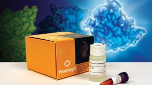
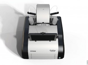
Promega kit depicted; test involves wavelength for a luciferase assay.

It’s a question I’m asked probably once a week. “What wavelength do I select on my luminometer when performing a luciferase assay?” The question is a good and not altogether unexpected one, especially for those new to bioluminescent assays. The answer is that in most cases, you don’t and in fact shouldn’t select a wavelength (the exception to this rule is if you’re measuring light emitted in two simultaneous luciferase reactions). To understand why requires a bit of an explanation of absorbance, fluorescence, and luminescence assays, and the differences among them.

Absorbance, fluorescence, and luminescence assays are all means to quantify something of interest, be that a genetic reporter, cell viability, cytotoxicity, apoptosis, or other markers. In principle, they are all similar. For example, a genetic reporter assay is an indicator of gene expression. The promoter of a gene of interest can be cloned upstream of a reporter such as β-galactosidase, GFP, or firefly luciferase. The amount of each of these reporters that is transcribed into mRNA and translated into protein by the cell is indicative of the endogenous expression of the gene of interest.

Continue reading “Why You Don’t Need to Select a Wavelength for a Luciferase Assay”

To inject or not inject?

GloMax® Discover Multimode Reader with injectors.

Luciferase assays are useful tools for studying a wide range of biological questions. They can be performed easily by adding a reagent that provides components necessary to generate a luminescent signal directly to cells or a cell lysate. However, once this reagent has been added, how long you wait to measure the signal becomes a key consideration in generating consistent data. Dependent on which luciferase assay you use, you may need a luminometer that can use injectors to deliver the assay reagents. The reason for this is simple, but can be confusing to new users.

Let’s start by discussing two types of luciferase assays: “flash” vs. “glow”. Continue reading “To inject or not inject?”

Optimize Your Western Blot

0447MA09_0A
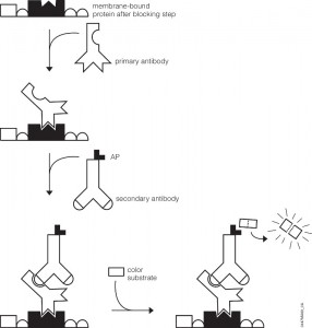
Western Blot Detection.

You’ve probably been there. You’ve got a new antibody or you’re testing out one you’ve made yourself. After weeks or months of work, your antibody is going to help move your research project forward. As you excitedly head to the dark room to develop your film, your mood is crushed when you see…bands, more bands, and smears. Alas, science has played one more cruel joke on you as you experience what so many of your fellow scientists have before. Despite such a dismal beginning, you often can still get good western blots by changing steps in your protocol.

Several steps in the western blot protocol can be optimized.

Continue reading “Optimize Your Western Blot”

Lessons From the ‘Long Goodbye’

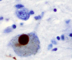
Lewy Body stained with alpha-synuclein.

A week ago Sunday, I walked among crowds of mothers, grandmothers, and children of all ages celebrating Mother’s Day at the Botanical Gardens in St. Louis, Missouri.  As I watched happy families, I couldn’t help being jealous.  Though I was there with my grandmother and other close relatives, I missed my mom, especially since I was in my hometown for her funeral the day before.  Had my mom been alive and well, we might have walked those same paths ourselves and enjoyed the new life teeming above the earth.  Instead, my mother lost her battle of more than six years with Lewy Body dementia the week before at the age of 61.

As a biologist, I was well-aware of Alzheimer disease in the abstract, and tau proteins, beta-amyloid, and genetic predisposition.  But until my mom was diagnosed in 2008, I was painfully ignorant of dementias other than Alzheimer disease.  Once we knew what mom was fighting, I learned that Alzheimer disease and Lewy Body are hardly unique.  The number of other dementias that exist is long and includes vascular dementia, mixed dementia, Parkinson’s disease, frontotemporal dementia, Creutzfeldt-Jakob disease, Huntington disease, and many others.[1]

Continue reading “Lessons From the ‘Long Goodbye’”

Lost in Translation? Tips for Preparing RNA for in vitro Translation Experiments

In vitro translation of proteins through cell-free expression systems using rabbit reticulocytes, E. coli S30, or wheat germ extracts can be invaluable in studying protein function.  If you only need a small amount (100s of nanograms), it’s also faster and easier than synthesizing vast quantities in bacterial or mammalian cells (~ 90 minutes for cell-free vs. long growth times and extraction steps after an initial optimization for protein synthesized in larger scale).  There are many systems out there, and knowing which to use can sometimes be difficult.  Many kits include components that combine transcription and translation in one-step, eliminating the need to provide your own RNA.  But when you want to make your own RNA templates to add to lysates, then there are additional concerns.

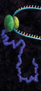
artists concept of in vitro translation
A protein chain being produced from a ribosome.

Many people don’t want to work with RNA since the common lab lore suggests it’s a finicky molecule, and for good reason.  Extracting it requires the utmost care in technique and elimination of nucleases.  Failing to do so results in degradation of the molecule, and so with it your experiments (see our recent blog by Terri Sundquist on tips for isolating RNA with ease).  Preparing RNA for cell-free expression is subject to the same concerns as extracted RNA, but with the proper care is not that much more of a challenge than using a DNA template.

The first step for using cell-free expression systems with RNA templates is to make the RNA.  Here are some tips that will ensure success.

Continue reading “Lost in Translation? Tips for Preparing RNA for in vitro Translation Experiments”