Some St. Patrick’s Day Science: Green Rivers, Four-Leaf Clovers and Optics of a Good Pint

St. Patrick’s Day means different things depending on where you are in the world. In Ireland, it’s a national holiday steeped in culture and tradition: parades, traditional music sessions and, for many, a pint of Guinness accompanied by a hearty “Sláinte” are all part of the day. Here in the American Midwest, we tend to turn that same spirit into a full spectacle. Green everything as far as the eye can see, including somehow an entire river.

Whatever your version of the holiday looks like, there is a lot of fun science behind it. Here is a look at Midwest St. Patrick’s Day through a lab lens.

The Chicago River: Where Orange Becomes Green

St. Patricks Day, Chicago River, Green

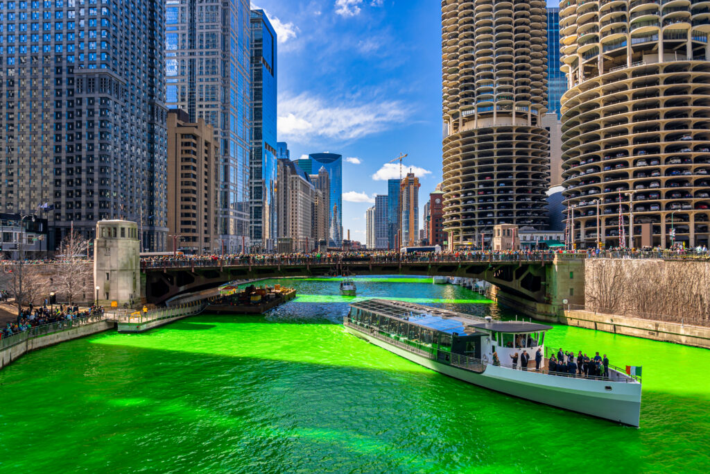
Every St. Patrick’s Day, the Chicago Journeymen Plumbers Local 130 heads out on the river and, in roughly 45 minutes, turns a stretch of the Chicago River a surreal emerald green (7, 11). The twist: the dye goes in orange.

The tradition dates to the early 1960s, rooted in a practical idea. Dye had been used to trace leaks and flow in the city’s waterways and someone realized the same concept could be repurposed into a public spectacle (7,11). The exact formula has been kept secret ever since, described only as environmentally friendly and designed to fade after a few hours (1,7,11).

Continue reading “Some St. Patrick’s Day Science: Green Rivers, Four-Leaf Clovers and Optics of a Good Pint”

Monochromator vs Filter-Based Plate Reader: Which is Better?

When it comes to purchasing a microplate reader for fluorescence detection, the most common question is whether to choose a monochromator-based reader or filter-based reader. In this blog, we’ll discuss how both types of plate readers work and factors to consider when determining the best plate reader for your need.

How do monochromator-based plate readers work?

Monochromators work by taking a light source and splitting the light to focus a particular wavelength on the sample. During excitation, the light passes through a narrow slit, directed by a series of mirrors and diffraction grating and then passes through a second narrow slit prior to reaching the sample. This ensures the desired wavelength is selected to excite the fluorophore. Once the fluorophore is excited, it emits light at a different, longer wavelength. This emission light is captured by another series of mirrors, grating and slits to limit the emission to a desired wavelength, which then enters a detector for signal readout.

Monochromator-based plate reader
Continue reading “Monochromator vs Filter-Based Plate Reader: Which is Better?”

Why You Don’t Need to Select a Wavelength for a Luciferase Assay

Promega kit depicted; test involves wavelength for a luciferase assay.

It’s a question I’m asked probably once a week. “What wavelength do I select on my luminometer when performing a luciferase assay?” The question is a good and not altogether unexpected one, especially for those new to bioluminescent assays. The answer is that in most cases, you don’t and in fact shouldn’t select a wavelength (the exception to this rule is if you’re measuring light emitted in two simultaneous luciferase reactions). To understand why requires a bit of an explanation of absorbance, fluorescence, and luminescence assays, and the differences among them.

Absorbance, fluorescence, and luminescence assays are all means to quantify something of interest, be that a genetic reporter, cell viability, cytotoxicity, apoptosis, or other markers. In principle, they are all similar. For example, a genetic reporter assay is an indicator of gene expression. The promoter of a gene of interest can be cloned upstream of a reporter such as β-galactosidase, GFP, or firefly luciferase. The amount of each of these reporters that is transcribed into mRNA and translated into protein by the cell is indicative of the endogenous expression of the gene of interest.

Continue reading “Why You Don’t Need to Select a Wavelength for a Luciferase Assay”