This blog is written by guest author, Maggie Bach, Sr. Product Manager, Promega Corporation.
Researchers are increasingly relying on cells grown in three-dimensional (3D) structures to help answer their research questions. Monolayer, or 2D cell culture, was the go-to cell culture method for the past century. Now, the need to better represent in vivo conditions is driving the adoption of 3D cell culture models. Cells grown in 3D structures better mimic tissue-like structures, better exhibit differentiated cellular functions, and better predict in vivo responses to drug treatment.
Switching to 3D cell culture models comes with challenges. Methods to interrogate these models need to be adaptable and reliable for the many types of 3D models. Some of the most popular 3D models include spheroids grown in ultra-low attachment plates, and cells grown in an extracellular matrix, such as Matrigel® from Corning. Even more complex models include medium flow over the cells in microfluidic or organ-on-a-chip devices. Will an assay originally developed for cells grown in monolayer perform consistently with various 3D models? How is measuring a cellular marker different when cells are grown in 3D models compared to monolayer growth?
Alternatives to animal testing have long been explored when it comes to studying the safety of various chemical compounds for use in food, medicine and cosmetics. With the advent of three-dimensional (3D) cell culture to create organoids, more relevant human organoid models are being explored as one way to provide safe and effective compound testing while minimizing the need for testing in animals. The international project Physiologically Anchored Tools for Realistic nanOmateriaL hazard aSsessment (PATROLS) led by the Swansea University Medical School aims to establish a battery of innovative, next-generation safety testing tools that can more accurately predict the effects of engineered nanomaterial (ENM) exposure in humans and the environment.
One project being researched by Samantha Llewellyn, a research assistant at Swansea University, is developing predictive physiologically relevant 3D liver models for ENM safety assessment. By having a model to evaluate realistic ENM exposures, a researcher can study liver function, hepatic metabolism and microtissue cell viability after acute (24 hours) or prolonged (several days) exposure. A microtissue model for assessing ENM hepatotoxicity needs to mimic primary hepatocytes and be amenable to assays used to test cell viability and metabolism.
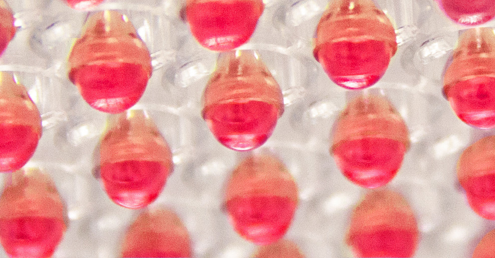
The right tools for testing this 3D liver model include the bioluminescent-based CellTiter-Glo® 3D Viability and P450-Glo® Assays. When creating organoids, having reagents that can penetrate to the center of the dense and complex 3D liver spheroids is important so that the cell viability readout encompasses the entire microtissue. The CellTiter-Glo® 3D Viability Assay accomplishes this task, providing accurate assessment of 3D tissue cell health. Measuring cytochrome P450 (CYP450) activity is necessary for studying liver function. The P450-Glo® Assays have the flexibility to assess CYP450 activity while preserving the liver spheroids; thus, researchers can gather more data from a single experiment.
The importance of Samantha Llewellyn’s research as part of PATROLs is establishing a 3D liver model that could evaluate realistic ENM exposures and reduce the need for animal testing. Bioluminescent assays for assessing cell health and liver enzyme function are necessary to reach this goal.
To learn more about the last 30 years of bioluminescent innovations and the discoveries they’ve enabled, please visit our 30th anniversary celebration page.
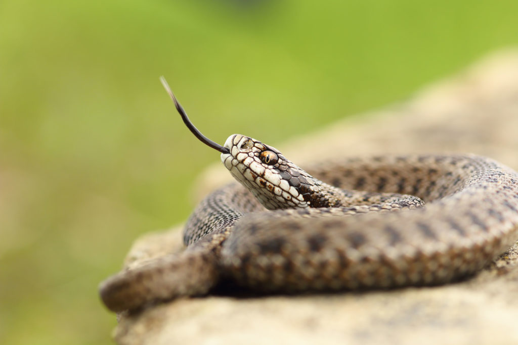
Snakebite is a serious public health issue in many tropical countries. Every year, roughly 2 million cases of poisoning from snakebites occur, and more than 100,000 people die. Snake venom is extremely complex, containing a cocktail of chemicals, many of which are undefined. This complicates the development of new therapeutics for treating snakebite.
Antivenom is the most effective treatment for snakebites,
but its production is complex and dangerous. It involves manually milking the
venom from different species of live snakes, then injecting small doses of the
venom into animals (mostly horses) to stimulate an immune response. After a
period of time, antibodies form in the animal’s blood, which is purified for
use as antivenom.
But what if we could produce snake venom in the lab, instead
of using live snakes? Recently, a group from the Netherlands did just that by
growing organoids derived from snake venom glands.
XWe use cookies and similar technologies to make our website work, run analytics, improve our website, and show you personalized content and advertising. Some of these cookies are essential for our website to work. For others, we won’t set them unless you accept them. To learn more about our approach to Privacy we invite you to Read More
By clicking “Accept All”, you consent to the use of ALL the cookies. However you may visit Cookie Settings to provide a controlled consent.
We use cookies and similar technologies to make our website work, run analytics, improve our website, and show you personalized content and advertising. Some of these cookies are essential for our website to work. For others, we won’t set them unless you accept them. To find out more about cookies and how to manage cookies, read our Cookie Policy.
If you are located in the EEA, the United Kingdom, or Switzerland, you can change your settings at any time by clicking Manage Cookie Consent in the footer of our website.
Necessary cookies are absolutely essential for the website to function properly. These cookies ensure basic functionalities and security features of the website, anonymously.
Cookie
Duration
Description
cookielawinfo-checbox-analytics
11 months
This cookie is set by GDPR Cookie Consent plugin. The cookie is used to store the user consent for the cookies in the category "Analytics".
cookielawinfo-checbox-functional
11 months
The cookie is set by GDPR cookie consent to record the user consent for the cookies in the category "Functional".
cookielawinfo-checbox-others
11 months
This cookie is set by GDPR Cookie Consent plugin. The cookie is used to store the user consent for the cookies in the category "Other.
cookielawinfo-checkbox-advertisement
1 year
The cookie is set by GDPR cookie consent to record the user consent for the cookies in the category "Advertisement".
cookielawinfo-checkbox-necessary
11 months
This cookie is set by GDPR Cookie Consent plugin. The cookies is used to store the user consent for the cookies in the category "Necessary".
cookielawinfo-checkbox-performance
11 months
This cookie is set by GDPR Cookie Consent plugin. The cookie is used to store the user consent for the cookies in the category "Performance".
gdpr_status
6 months 2 days
This cookie is set by the provider Media.net. This cookie is used to check the status whether the user has accepted the cookie consent box. It also helps in not showing the cookie consent box upon re-entry to the website.
lang
This cookie is used to store the language preferences of a user to serve up content in that stored language the next time user visit the website.
viewed_cookie_policy
11 months
The cookie is set by the GDPR Cookie Consent plugin and is used to store whether or not user has consented to the use of cookies. It does not store any personal data.
Analytical cookies are used to understand how visitors interact with the website. These cookies help provide information on metrics the number of visitors, bounce rate, traffic source, etc.
Cookie
Duration
Description
SC_ANALYTICS_GLOBAL_COOKIE
10 years
This cookie is associated with Sitecore content and personalization. This cookie is used to identify the repeat visit from a single user. Sitecore will send a persistent session cookie to the web client.
vuid
2 years
This domain of this cookie is owned by Vimeo. This cookie is used by vimeo to collect tracking information. It sets a unique ID to embed videos to the website.
WMF-Last-Access
1 month 18 hours 24 minutes
This cookie is used to calculate unique devices accessing the website.
_ga
2 years
This cookie is installed by Google Analytics. The cookie is used to calculate visitor, session, campaign data and keep track of site usage for the site's analytics report. The cookies store information anonymously and assign a randomly generated number to identify unique visitors.
_gid
1 day
This cookie is installed by Google Analytics. The cookie is used to store information of how visitors use a website and helps in creating an analytics report of how the website is doing. The data collected including the number visitors, the source where they have come from, and the pages visted in an anonymous form.
Advertisement cookies are used to provide visitors with relevant ads and marketing campaigns. These cookies track visitors across websites and collect information to provide customized ads.
Cookie
Duration
Description
IDE
1 year 24 days
Used by Google DoubleClick and stores information about how the user uses the website and any other advertisement before visiting the website. This is used to present users with ads that are relevant to them according to the user profile.
test_cookie
15 minutes
This cookie is set by doubleclick.net. The purpose of the cookie is to determine if the user's browser supports cookies.
VISITOR_INFO1_LIVE
5 months 27 days
This cookie is set by Youtube. Used to track the information of the embedded YouTube videos on a website.
Performance cookies are used to understand and analyze the key performance indexes of the website which helps in delivering a better user experience for the visitors.
Cookie
Duration
Description
YSC
session
This cookies is set by Youtube and is used to track the views of embedded videos.
_gat_UA-62336821-1
1 minute
This is a pattern type cookie set by Google Analytics, where the pattern element on the name contains the unique identity number of the account or website it relates to. It appears to be a variation of the _gat cookie which is used to limit the amount of data recorded by Google on high traffic volume websites.