From Chromosomes to the Brain: How HaloTag Is Expanding What We Can See

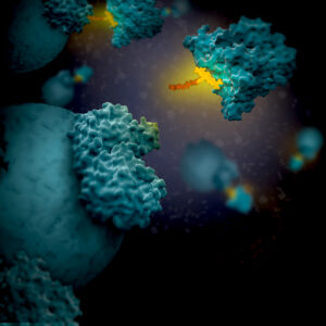
Artist's 3D concept of HaloTag with linker attaching to a target protein

Sometimes in the life sciences, it’s really tempting to look at a process like mitosis and think, “Wow, we really understand that,” and stop asking questions. In other cases, significant barriers like reliable expression of a non-endogenous reporter in an organ such as the brain is a challenge that seems impossible to overcome. However, those are precisely the moments when science and scientists need to push the boundaries of what we can see. In this post, I discuss two studies where researchers are literally seeing what could not be seen before by developing new methods for looking at their model systems.

In both papers discussed here, the HaloTag® protein and ligands were key to developing new methods allowing scientists to visualize biological phenomena that had been previously inaccessible. HaloTag® protein allows different functionalities to be linked onto a single genetic construct, rapidly and covalently under physiological conditions for experiments ranging from live-cell imaging to capture of protein interaction complexes (1). In the first study, HaloTag-CENP-A + JF635 ligand was the specific tool that let them directly see, in real time, that chromosome-attached microtubules pivot as the spindle elongates. In the second study, HaloTag was used to create a protocol for PET imaging of reporter gene systems in the brain.

Continue reading “From Chromosomes to the Brain: How HaloTag Is Expanding What We Can See”

An Unexpected Role for RNA Methylation in Mitosis Leads to New Understanding of Neurodevelopmental Disorders

Traditionally, RNA methylation has been studied in the context of gene expression regulation, RNA stability and translation efficiency, with its primary role thought to be in modulating cellular homeostasis and protein synthesis. However, a 2025 study by Dharmadkikari and colleagues uncovers an unexpected and critical function for RNA methylation in mitotic spindle integrity.

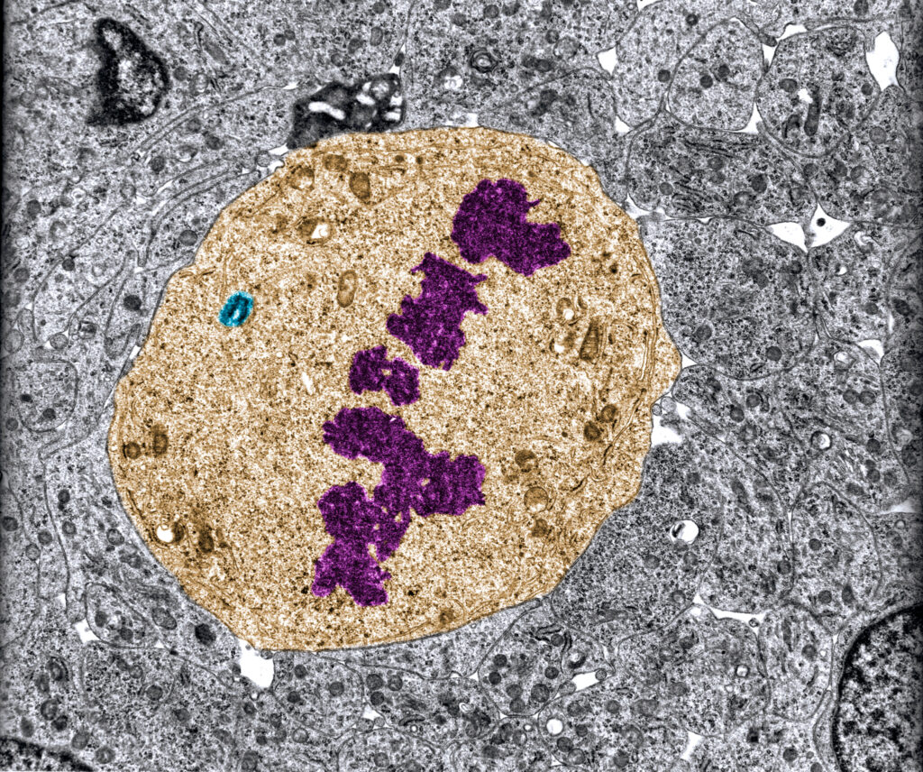
False color transmission electron microscope (TEM) micrograph of a mitotic cell in metaphase stage showing chromosomes (purple) in the equatorial plane and one of the mitotic spindle poles (blue). Mutations in SPOUT1/CENP-32 affect RNA methylation which is necessary for proper cell division.
False color transmission electron microscope (TEM) micrograph of a mitotic cell in metaphase stage showing chromosomes (purple) in the equatorial plane and one of the mitotic spindle poles (blue).

The study identifies a critical role for SPOUT1/CENP-32-dependent methylation in mitotic spindle formation and accurate chromosome segregation. Originally identified in a large-scale analysis of proteins associated with mitotic chromosomes, SPOUT1/CENP-32 encodes a putative RNA methyltransferase. The protein localizes to mitotic spindles, and when it is absent centrosome detachment from the spindle poles, delayed anaphase, and chromosome segregation errors are observed. Further, CRISPR experiments in human cells show that the protein is essential for cell viability.

Continue reading “An Unexpected Role for RNA Methylation in Mitosis Leads to New Understanding of Neurodevelopmental Disorders”