Advancing Understanding of Hypoxic Gene Regulation Using Reporter Genes: Celebrating the Work of Dr. Gregg L. Semenza

This post is written by guest blogger, Amy Landreman, PhD, Sr. Product Manager at Promega Corporation.

Oxygen is necessary for animal life. It’s essential for cellular respiration and the production of energy (ATP) we require to survive. Given the need for oxygen, it isn’t surprising that our bodies have evolved ways to sense and adapt to decreased oxygen conditions (hypoxia). We can increase the production of new blood vessels by producing vascular endothelial growth factor (VEGF) or increase red blood cell (RBC) production by increasing the levels of eythropoietin (EPO), the hormone that plays a key role in the production of RBCs. But how does our body sense low oxygen, increase EPO levels, and kick our RBC production into gear? Nobel laureate Gregg L. Semenza has been honored for his contributions to our understanding of this process, and his research demonstrates the value of reporter genes and bioluminescence for studying gene regulation.
Reporter genes and bioluminescence are important tools for studying gene regulation
Continue reading “Advancing Understanding of Hypoxic Gene Regulation Using Reporter Genes: Celebrating the Work of Dr. Gregg L. Semenza”

Looking Back: Cell-Free Expression Systems Helped to Characterize Proteins Involved in Hypoxia Response

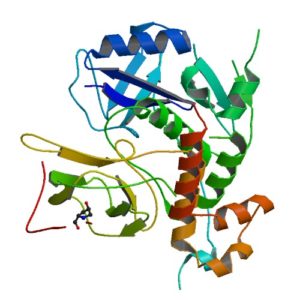
Structur of a HIF-1a-pVHL-ElonginB-ElonginC complex
Structure of a HIF-1a-pVHL-ElonginB-ElonginC complex

William G. Kaelin Jr., Sir Peter J. Ratcliffe and Gregg L. Semenza were awarded the 2019 Nobel Prize in Physiology or Medicine for their discoveries of how cells sense and adapt to oxygen availability.

Kaelin and Ratcliffe’s labs focused their efforts on the transcription factor HIF (hypoxia-inducible factor). This transcription factor is critical in the cellular adaptation of to changes in oxygen availability.

When oxygen levels are elevated cells contain very little HIF. Ubiquitin is added to the HIF protein via the VHL complex and it is degraded in the proteasome.  When oxygen levels are low (hypoxia) the amount of HIF increases.

In 2001 both groups published articles characterizing the interaction between VHL and HIF, and these articles were referenced by the Nobel Prize Organization in their press release about this year’s award. (1,2). Both studies demonstrated that under the normal oxygen conditions hydroxylation of proline residue P564 enabled VHL to recognize and bind to HIF.

The use of cell free expression (i.e., TNT Coupled Transcription/Translation System) by both labs was key in the characterization of the VHL:HIF interaction The labs utilized HIF and VHL 35-S labeled proteins generated via the TNT system under both normal or in a hypoxic work station to:

  • Determine the affect of ferrous chloride and cobaltous chloride on the interaction
  • Map the specific region of HIF required for the interaction to occur (556-574)
  • Determine the effect of HIF point mutations on the interaction
  • Use synthetic peptides to block the interaction
  • Conclude that a factor in mammalian cells was necessary for the interaction to occur.

Literature Cited

  1. Ivan, M et al. (2001) HIF Targeted for VHL-Mediated Destruction by Proline Hydroxylation: Implications for O2 Sensing. Science 292: 464–67.
  2. Jaakkola, P. et al. (2001) Targeting of HIF-α to the von Hippel-Lindau Ubiquitylation of Complex by O2– Regulated Prolyl Hydroxylation. Science 202, 468–72 .

Related Posts

How Autophagy Feeds Cancer’s Need for Metabolites

Illustration of energy metablism in cell.

Metabolism underpins numerous cellular processes. Without it, cells would not grow, divide, synthesize or secrete. Another pathway, autophagy, degrades unwanted cellular materials, helping to maintain cell health. With these opposing roles, is there a connection between autophagy and metabolism? As it turns out, the answer is yes. Because molecules degraded by autophagy are recycled and fed into metabolism pathways as precursor compounds. There are interesting implications as a result of this connection, ones that affect cancer cells as described in a recent Cell Metabolism review article.

Autophagic flux, the process by which molecules and organelles are directed to the autophagosome, fuse with the lysosome and are degraded, involves a selective process that determines the cargo carried within the autophagosome. Autophagy-related genes (ATGs) direct the process and particular receptor proteins bind the cargo. What is interesting about the connection among cancer, autophagy and metabolism is the complexity of the role that autophagy plays in cancer. While autophagy was thought to act in a more tumor suppressive manner as shown when one copy of an ATG6 analogous gene in mice was deleted and the other left unaltered, and malignant tumors developed, but in mice mosaic for ATG5 deletions, the inhibition of autophagy resulted in benign tumors in the liver. This latter experiment suggested autophagy was needed for cancer progression, a hypothesis reinforced by the lack of ATG mutations in human cancers.

Continue reading “How Autophagy Feeds Cancer’s Need for Metabolites”