Immunometabolism is the study of how metabolic processes influence immune cell functions and how immune responses, in turn, shape cellular metabolism. This field examines the roles of cytokines and metabolites, which act as signaling molecules and energy sources, respectively. Cytokines can trigger or modulate metabolic pathways in immune cells, affecting their activation, growth, and response capabilities. Similarly, metabolites provide the necessary energy and building blocks that enable immune cells to proliferate, function optimally, and sustain their activity during immune responses. This dynamic interplay is crucial for maintaining health and combating disease. Together, cytokines and metabolites orchestrate a complex network that links metabolic health with immune competence on a systemic and cellular level. This blog discusses how cytokines and metabolites not only influence but also drive immune cell functions, revealing new avenues for therapeutic interventions across a range of diseases.
In the past decade, there has been a sharp rise in studies using spheroids as cell models for basic research and drug discovery. Spheroids are self-organized aggregation of cells that form a spherical mass, and they have become widely popular because they are much more physiologically relevant compared to flat 2D cell cultures.
In spheroids, the inner cells have less access to nutrients and oxygen compared to the outer layer, forming a natural gradient. As a result, metabolite concentration and cellular state such as proliferation and differentiation, can be very different at the periphery compared to the inner core. This phenomenon, known as “heterogeneity”, makes 3D tumor spheroids much more representative of actual tumors in the human body.
While you can rely on Taylor Swift and Adele to help heal emotional heartbreak, unfortunately treating a physically “broken” heart, a heart damaged by fibrosis, is a much more complicated process than putting on your favorite sad songs and wallowing in your feelings. In a recent study published in Science, researchers developed a therapeutic approach to treat damaged hearts in mice through the removal of scar tissue using genetically engineered immune cells (CAR T cells) and the mRNA technology used in the mRNA coronavirus vaccines.
Electrophysiology experiments provide a view into the cell with amazing detail. The paper reviewed here describes a molecular reporter biosensor (NanoBRET) that can offer the same kind of temporal and spatial resolution traditionally reserved for extremely labor-intensive experiments like patch clamp analysis.
I confess that I struggled through biophysics, and my Bertil Hille textbook Ion Channels of Excitable Membranes lies neglected somewhere in a box in my basement (I have not tossed it into the recycle bin—I can’t bear too, I spent too much time bonding with that book in graduate school).
My struggles in that graduate class and my attendance at the seminars of my grad school colleagues who were conducting electrophysiological studies left me with a sincere awe and appreciation of both the genius and the artistry required to produce nice electrophysiology data. The people who are good at these experiments are artists—they have the golden touch when it comes to generating that megaohm seal between a piece of cell membrane and a finely pulled glass pipette. And, they are brilliant scientists, they really understand the physics, the chemistry and the biology of the cells they study from a perspective that very few scientists ever develop.
Electrophysiology data, which often demonstrate the gating of a single channel protein in response to a single stimulus in real time–ions crossing a membrane through a single protein–are amazing for their ability, unlike virtually any other experimental data for the story they can tell about what is going on in a cell in real time under physiological conditions.
When constructs were ectopically expressed in HEK 293T/17 cells, we obtained very similar kinetics for the GPCR-driven responses between NanoBRET™ biosensors and the patch clamp recordings.
They continue:
Indeed, the activation rates that we observed were very similar to those of GPCR-stimulated GIRKs [G protein-coupled, inwardly rectifying K+ channel] in native cells, suggesting that the conditions of this assay closely match the in vivo setting. This finding further demonstrates the ability of the system to resolve the fast, physiological relevant kinetics of GPCR signaling.
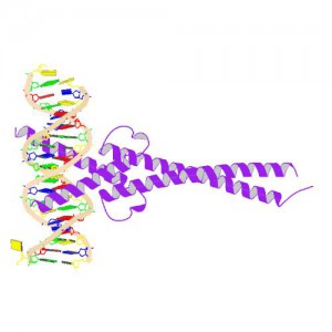
Crystal Structure of MYC MAX Heterodimer bound to DNA ImageSource=RCSB PDB; StructureID=1nkp; DOI=http://dx.doi.org/10.2210/pdb1nkp/pdb;
In 1982, picked up because of its homology to chicken virus genes that could transform cells, MYC became one of the first human genes identified that could drive cellular transformation (1,2). Since that time countless laboratories have prodded and poked the human MYC gene, the MYC protein, their homologs in other animal models, and their transforming viral counterparts.
MYC is a transcription factor and forms heterodimers with a required protein partner, MAX, before binding to the E box sequences of DNA regulatory regions (3). MYC regulates gene expression of many targets through interactions with a host of proteins, often referred to as the MYC Interactome (2). In fact, MYC is estimated to bind 10–15% of the genome, and it regulates the expression of genes that are transcribed by by each of the three RNA polymerases (2).
MYC plays a central role in regulating cell growth, proliferation, apoptosis, differentiation and transformation, acting as a central integrator of cellular signals. MYC is tightly regulated at multiple levels from gene expression to protein stability. Dysregulation (usually upregulation) of the amount and stability of Myc protein is observed in many human cancers. Even in cancers in which MYC is not directly involved in transforming cells, its normal expression is often required to support the extracellular matrix and/or vascularization necessary for tumor growth and formation (4).
Because MYC is such a central player cancer pathology, it is an attractive target for cancer therapeutics (2) .
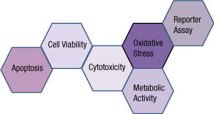
You often need several pieces of information to really understand what is happening within a cell or population of cells. If your cells are not proliferating, are they dying? Or, are you seeing cytostasis? If they are dying, what is the mechanism? Is it apoptosis or necrosis? If you are seeing apoptosis, what is the pathway: intrinsic or extrinsic?
If you are measuring expression of a reporter gene and you see a decrease in expression, is that decrease due to transfection inefficiencies, cytotoxicity, or true down regulation of your reporter gene?
To investigate these multiple parameters, you can run assays in parallel, but that requires more sample, and sample isn’t always abundant.
Multiplexing assays allows you to obtain information about multiple parameters or events (e.g., reporter gene expression and cell viability; caspase-3 activity and cell viability) from a single sample. Multiplexing saves sample, saves time and gives you a more complete picture of the biology that is happening with your experimental sample.
What information do you need about your cells to complete the picture?
Multiplexing assay reagents to measure biomarkers in the same sample has often been considered an application only accomplished with antibodies or dyes and sophisticated detection instrumentation. However, Promega has developed microwell plate based assays for cells in culture that allow multiplexed detection of biomarkers in the same sample well using standard multimode multiwell plate readers. Continue reading “Piecing the Puzzle Together: Using Multiple Assays to Better Understand What Is Happening with Your Cells”
XWe use cookies and similar technologies to make our website work, run analytics, improve our website, and show you personalized content and advertising. Some of these cookies are essential for our website to work. For others, we won’t set them unless you accept them. To learn more about our approach to Privacy we invite you to Read More
By clicking “Accept All”, you consent to the use of ALL the cookies. However you may visit Cookie Settings to provide a controlled consent.
We use cookies and similar technologies to make our website work, run analytics, improve our website, and show you personalized content and advertising. Some of these cookies are essential for our website to work. For others, we won’t set them unless you accept them. To find out more about cookies and how to manage cookies, read our Cookie Policy.
If you are located in the EEA, the United Kingdom, or Switzerland, you can change your settings at any time by clicking Manage Cookie Consent in the footer of our website.
Necessary cookies are absolutely essential for the website to function properly. These cookies ensure basic functionalities and security features of the website, anonymously.
Cookie
Duration
Description
cookielawinfo-checbox-analytics
11 months
This cookie is set by GDPR Cookie Consent plugin. The cookie is used to store the user consent for the cookies in the category "Analytics".
cookielawinfo-checbox-functional
11 months
The cookie is set by GDPR cookie consent to record the user consent for the cookies in the category "Functional".
cookielawinfo-checbox-others
11 months
This cookie is set by GDPR Cookie Consent plugin. The cookie is used to store the user consent for the cookies in the category "Other.
cookielawinfo-checkbox-advertisement
1 year
The cookie is set by GDPR cookie consent to record the user consent for the cookies in the category "Advertisement".
cookielawinfo-checkbox-necessary
11 months
This cookie is set by GDPR Cookie Consent plugin. The cookies is used to store the user consent for the cookies in the category "Necessary".
cookielawinfo-checkbox-performance
11 months
This cookie is set by GDPR Cookie Consent plugin. The cookie is used to store the user consent for the cookies in the category "Performance".
gdpr_status
6 months 2 days
This cookie is set by the provider Media.net. This cookie is used to check the status whether the user has accepted the cookie consent box. It also helps in not showing the cookie consent box upon re-entry to the website.
lang
This cookie is used to store the language preferences of a user to serve up content in that stored language the next time user visit the website.
viewed_cookie_policy
11 months
The cookie is set by the GDPR Cookie Consent plugin and is used to store whether or not user has consented to the use of cookies. It does not store any personal data.
Analytical cookies are used to understand how visitors interact with the website. These cookies help provide information on metrics the number of visitors, bounce rate, traffic source, etc.
Cookie
Duration
Description
SC_ANALYTICS_GLOBAL_COOKIE
10 years
This cookie is associated with Sitecore content and personalization. This cookie is used to identify the repeat visit from a single user. Sitecore will send a persistent session cookie to the web client.
vuid
2 years
This domain of this cookie is owned by Vimeo. This cookie is used by vimeo to collect tracking information. It sets a unique ID to embed videos to the website.
WMF-Last-Access
1 month 18 hours 24 minutes
This cookie is used to calculate unique devices accessing the website.
_ga
2 years
This cookie is installed by Google Analytics. The cookie is used to calculate visitor, session, campaign data and keep track of site usage for the site's analytics report. The cookies store information anonymously and assign a randomly generated number to identify unique visitors.
_gid
1 day
This cookie is installed by Google Analytics. The cookie is used to store information of how visitors use a website and helps in creating an analytics report of how the website is doing. The data collected including the number visitors, the source where they have come from, and the pages visted in an anonymous form.
Advertisement cookies are used to provide visitors with relevant ads and marketing campaigns. These cookies track visitors across websites and collect information to provide customized ads.
Cookie
Duration
Description
IDE
1 year 24 days
Used by Google DoubleClick and stores information about how the user uses the website and any other advertisement before visiting the website. This is used to present users with ads that are relevant to them according to the user profile.
test_cookie
15 minutes
This cookie is set by doubleclick.net. The purpose of the cookie is to determine if the user's browser supports cookies.
VISITOR_INFO1_LIVE
5 months 27 days
This cookie is set by Youtube. Used to track the information of the embedded YouTube videos on a website.
Performance cookies are used to understand and analyze the key performance indexes of the website which helps in delivering a better user experience for the visitors.
Cookie
Duration
Description
YSC
session
This cookies is set by Youtube and is used to track the views of embedded videos.
_gat_UA-62336821-1
1 minute
This is a pattern type cookie set by Google Analytics, where the pattern element on the name contains the unique identity number of the account or website it relates to. It appears to be a variation of the _gat cookie which is used to limit the amount of data recorded by Google on high traffic volume websites.