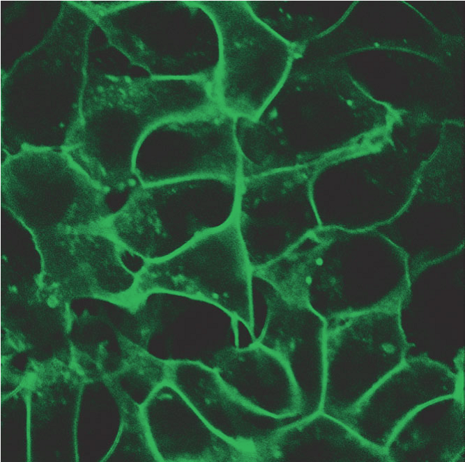
Cell signaling is a finely tuned process where both timing and spatial context play essential roles. Whether it’s a hormone triggering a cellular response or a drug modulating a pathway, these processes unfold in dynamic, spatially organized ways. To study them, researchers rely on chemigenetic biosensors—genetically encoded tools that light up in response to molecular activity. However, traditional biosensors are constrained by several limitations: poor photostability under prolonged imaging, limited spectral flexibility for multiplexing, and insufficient spatial resolution for studying signaling events at subcellular scales.
Introducing HaloAKARs: A New Class of Biosensors
A recent study published in Nature Biotechnology introduces a new class of far-red chemigenetic biosensors—called HaloAKARs—that may overcome the limitations of existing biosensors. HaloAKARs combine self-labeling protein tags (HaloTag) with synthetic far-red fluorophores to achieve high-sensitivity, high-resolution imaging of kinase activity in living cells.
The core of the HaloAKAR design centers around a biosensor that detects the activity of protein kinase A (PKA), a key player in cellular responses to cAMP. The researchers engineered a sensing domain that undergoes a conformational change upon phosphorylation by PKA, which in turn alters the fluorescence intensity of the attached JF635 fluorophore. Through a high-throughput engineering approach, they screened over 15,000 biosensor variants to identify optimal versions. The standout HaloAKAR variant exhibited an impressive dynamic range—over a 12-fold change in fluorescence upon activation, which surpasses all existing PKA biosensors in sensitivity.
Super-Resolution Imaging and Subcellular Precision
What truly sets HaloAKARs apart is their compatibility with advanced imaging techniques. Using stimulated emission depletion (STED) microscopy, the researchers were able to visualize PKA activity at individual clathrin-coated pits, submicron structures involved in endocytosis. This level of resolution revealed a surprising heterogeneity—even neighboring pits could exhibit markedly different levels of kinase activity. This demonstrates the power of coupling biosensors with super-resolution imaging to uncover new biological insights.
Beyond PKA, the modular architecture of these biosensors allows them to be adapted for other kinases. By swapping out substrate peptides and recognition domains, the team created new sensors for PKC, Akt, and ERK, all of which performed robustly in live-cell imaging. These sensors were also effective in three-dimensional systems, including spheroids and primary pancreatic islets, showing that the technology is well-suited for physiologically relevant models.
Multiplexing Signaling Readouts in Real Time
A particularly exciting feature of the HaloAKAR platform is its capacity for multiplexing—imaging multiple biosensors at once without spectral overlap. Traditional fluorescent proteins tend to crowd the visible spectrum, but the far-red emission of HaloAKARs allows researchers to add more sensors into the mix. In one example, the team imaged five different signaling molecules—Ca2+, cAMP, cGMP, PKC activity, and PKA activity—within the same cell. This approach opens the door to exploring how complex signaling networks are orchestrated and how they differ across cell types, conditions, or drug treatments.
The authors applied this technique to study signaling downstream of various G-protein-coupled receptors (GPCRs). Their data revealed receptor-specific signaling patterns, including previously unrecognized cross-talk between G protein pathways. For example, stimulation of the GLP-1 receptor induced a sequence of rapid calcium and PKC signals followed by slower, sustained cAMP and PKA activation. These dynamics may explain the receptor’s physiological role in insulin secretion and its therapeutic relevance in diabetes and obesity.
The Future of Imaging
In summary, HaloAKARs combine spectral orthogonality, high sensitivity, and adaptability across multiple kinase pathways, providing a powerful tool for studying cellular signaling with unprecedented resolution. It not only enables scientists to see more, but to see more clearly—tracking signaling as it unfolds in real time, in real space, and across complex molecular landscapes. In the future, these new biosensors may help us have a more integrated view of how cells make decisions.
Reference: Frei, M.S. et al. (2025) Far-red chemigenetic kinase biosensors enable multiplexed and super-resolved imaging of signaling networks. Nat. Biotechnol., in press.
