Detecting Neuroinflammation in Microglia and Astrocytes

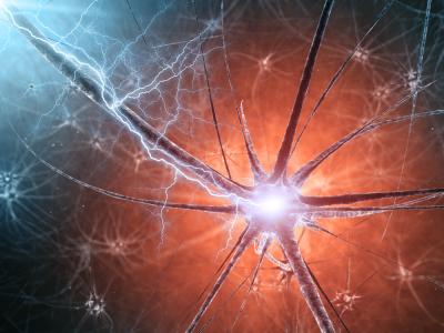
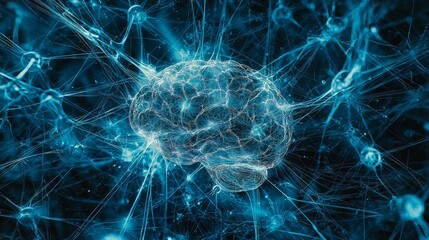
The brain is one of the most complex and fascinating parts of biology. Thankfully, it’s also remarkably good at protecting itself. When exposed to a pathogen, an injury or even misfolded proteins, microglia and astrocytes function as the central nervous system’s (CNS) primary immune defenders. They mount an inflammatory response by releasing cytokines and working to contain the damage. Yet this same system can malfunction or not resolve, which manifests as devastating consequences.

Chronic neuroinflammation is now recognized as a shared characteristic across some of the most common and difficult-to-treat neurological conditions. A 2023 review in Signal Transduction and Targeted Therapy highlighted the dualistic nature of neuroinflammation: while acute responses serve a protective role, chronic or dysregulated inflammatory signaling can initiate and accelerate neurodegeneration, identifying these pathways as priority targets for therapeutic intervention (Zhang et al., 2023). A 2025 review in Science reinforced this view, noting that within Multiple Sclerosis, disease-modifying therapies targeting neuroinflammation have seen the most clinical success (Shi & Yong, 2025). This could suggest applications within neurological conditions where the same inflammatory mechanisms are at work.

Understanding how and where these inflammatory signals originate in the CNS is an active area of preclinical research. One cytokine being actively studied is IL-6. IL-6 is produced by several cell types, including astrocytes and microglia in the CNS. As a key mediator of inflammatory responses, it mediates pro-inflammatory effects through its trans-signaling, which occurs via soluble IL-6 receptors. Dysregulation of this mechanism may contribute to the chronic neuroinflammation seen in several neurological conditions. Characterizing how and when IL-6 is secreted from CNS cells is an important step toward understanding the neuroinflammatory processes underlying these disorders.

Continue reading “Detecting Neuroinflammation in Microglia and Astrocytes”

Light Has a Favorite Color, But It’s Complicated

Last spring, my niece and I made a trip to a home improvement store to put together a Mother’s Day planter for my sister. My niece had a clear vision: my sister’s favorite color is blue, so we were going to buy blue flowers. We walked every aisle of the garden center. We checked the annuals, the perennials, and the hanging baskets then left with purple, red, and a grumpy 7-year-old.

It turns out we were not up against a bad selection. We were up against biology.

The Problem with Blue

Blue is one of the rarest colors in the natural world. The food industry is currently finding that out the hard way. There is a good chance you have eaten something blue today. Maybe it was the frosting on a birthday cake, the coating on some M&M’s® candies, or the sports drink in your refrigerator. That blue almost certainly came from a petroleum-based synthetic dye, and for the first time in decades, the food industry is being asked to find something better.

The FDA banned Red Dye No. 3 in January 2025, and pressure has been building around the remaining synthetic dyes ever since, including Blue No. 1 and Blue No. 2. Major food brands have begun announcing plans to reformulate.

There is just one problem. Blue is genuinely, stubbornly hard to make in nature. It turns out that blue has almost nothing to do with color, and almost everything to do with light.

Continue reading “Light Has a Favorite Color, But It’s Complicated”

Why BRETSA™ Target Engagement Matters for Drug Discovery

Drug discovery researchers face a fundamental constraint in their work to develop safe, effective therapeutics: the vast majority of the human proteome remains inaccessible to conventional small molecule approaches. Proteins without defined binding pockets, those lacking known chemical probes, and protein targets that fail to translate from biochemical assays into cellular models have long been considered out of reach of standard drug discovery screening tools. As Dixit et al. describe, developing biochemical or cellular assays for all genome-encoded targets “is not scalable and likely impossible as most proteins have ill-defined or unknown activity” — these are what the authors call “the dark undruggable expanses” of the proteome [1].

That gap is now narrowing. Promega Corporation recently launched the TarSeer™ BRETSA™ Target Engagement System, a live-cell target engagement platform designed to bring previously challenging targets within reach of early-stage drug discovery.

The Problem: A Translation Gap in Early Discovery

Drug discovery teams regularly encounter a frustrating disconnect. A compound may show strong binding activity in a biochemical assay, only to fail when tested in a cellular environment. Without target-specific cellular assays, which generally aren’t available for poorly characterized proteins, researchers face difficult choices when deciding which compounds to advance through the drug development pipeline.

Continue reading “Why BRETSA™ Target Engagement Matters for Drug Discovery”

Trends and Tools Transforming Drug Discovery: Five Takeaways from Discover Glo 2025

In biologics, cell therapy, and targeted protein degradation, the science is moving fast—and so are the tools. From GPCR-targeted therapies to real-time CAR-T manufacturing tools, new techniques are reshaping how scientists approach drug development, live-cell imaging, and protein degradation.

The “Bringing Light to Science” Discover Glo 2025 speaker series brought together researchers from across academia and industry to share real-world examples of how bioluminescent technologies are helping them advance their research. Now available on demand, these sessions offer fresh perspectives and actionable takeaways on the future of therapeutic development, cellular analysis and assay design.

We’ve distilled five key takeaways from the sessions—practical insights you can apply to your own work or use to stay current with where the field is heading.

Continue reading “Trends and Tools Transforming Drug Discovery: Five Takeaways from Discover Glo 2025”

A New View of Protein Degradation with HiBiT and Live Cell Imaging

Targeted protein degradation (TPD) is an emerging drug discovery strategy that offers an entirely different approach to tackle disease-relevant proteins, including classic “undruggable” targets. Instead of inhibiting protein function, small molecules like PROTACs and molecular glues co-opt the cell’s own ubiquitin-proteasome system to eliminate specific proteins altogether. But as this targeted approach gains traction, it also challenges existing methods for validating compound activity.  

How do you confirm that degradation is happening in a biologically-relevant system? Can you validate protein degradation in real-time?  

Continue reading “A New View of Protein Degradation with HiBiT and Live Cell Imaging”

Understanding Wnt Signaling Through β-Catenin Localization in Live Cells

The Wnt/β-catenin pathway, long studied in the context of developmental biology, has become increasingly recognized for its role in a wide range of human diseases. Its dysregulation has been implicated in cancer, fibrosis, immune modulation, and neurodegenerative conditions—making it a clinically actionable target across diverse therapeutic areas1. In this blog, we cover the fundamentals of Wnt/β-catenin signaling, highlight ongoing research efforts to understand its role in disease, and show how combining live-cell imaging with luminescent assays complements functional studies.

Continue reading “Understanding Wnt Signaling Through β-Catenin Localization in Live Cells”

Using Dual-Luciferase Assays to Identify the Role of Non-Coding RNAs in Disease

In recent years, non-coding RNAs—especially microRNAs (miRNAs) and long non-coding RNAs (lncRNAs)—have emerged as powerful regulators of cellular behavior. These molecules modulate gene expression, often by targeting mRNAs for translational suppression or degradation. Two recent studies—one focused on osteoarthritis and the other on 5-Fu-resistant colorectal cancer—illustrate how these non-coding, regulatory RNAs operate within disease-relevant signaling networks, providing new points for therapeutic intervention.

lncRNA, long noncoding RNA

Both studies use the pmirGLO Dual-Luciferase miRNA Target Expression Vector to evaluation predicted miRNA activity.  This dual-luciferase system offers a clean and quantifiable way to validate miRNA–mRNA interactions using a simple bioluminescent readout. By cloning the 3´ untranslated regions (UTRs) of suspected targets downstream of a firefly luciferase reporter and normalizing against Renilla luciferase, researchers can rapidly confirm whether a miRNA directly regulates its target.

Continue reading “Using Dual-Luciferase Assays to Identify the Role of Non-Coding RNAs in Disease”

Bioluminescence vs. Fluorescence: Choosing the Right Assay for Your Experiment 

From enzyme activity to gene expression, light-based assays have become foundational tools in life science research. Among these, fluorescence and bioluminescence are two of the most widely-used approaches for detecting and quantifying biological events. Both rely on the emission of light, but the mechanisms generating that light—and the practical implications for experimental design—are quite different. 

Choosing between a fluorescence or bioluminescence assay isn’t as simple as picking between two reagents off the shelf. Each has strengths and limitations depending on the application, instrumentation, and biological system. In this blog, we’ll walk through how each method works, where they shine (and where they don’t), and what to consider when deciding which approach is right for your experiment. 

Continue reading “Bioluminescence vs. Fluorescence: Choosing the Right Assay for Your Experiment “

Advancing Neurodegenerative Disease Modeling: A Novel iPSC-Based Luminescence System for Parkinson’s Disease Research

Advancing our understanding of neurodegenerative diseases requires model systems that faithfully recapitulate the biology of human neurons. A recent study by Gandy et al. in the International Journal of Molecular Sciences introduces an innovative luminescence-based platform to explore the role of Parkinson’s disease (PD)-associated genes in living cells. By leveraging human induced pluripotent stem cells (iPSCs) and CRISPR-mediated endogenous tagging, researchers at the Early Drug Discovery Unit at The Neuro (Montreal Neurological Institute-Hospital) at McGill University and Health Canada have created a powerful system for investigating protein expression and function in a physiologically relevant setting.

Continue reading “Advancing Neurodegenerative Disease Modeling: A Novel iPSC-Based Luminescence System for Parkinson’s Disease Research”

Alzheimer Disease and Metabolic Dysfunction: A Critical Intersection in Brain Health

This guest blog post is written by Alden Little, a Marketing Intern at Promega.

Alzheimer disease (AD) is one of the most devastating neurodegenerative disorders, affecting millions worldwide. While much attention has been given to amyloid plaques and tau tangles, emerging research suggests that metabolic dysfunction in the brain plays a crucial role in the disease’s progression. A recent study published in Acta Neuropathologica by Schröder et al. sheds new light on how astrocytes—the brain’s metabolic support cells—are affected in AD, and how their dysfunction impacts neurons.

Auguste Deter, a patient of Dr. Alzheimer, who first described the hallmark plaques and tangles of AD.
Continue reading “Alzheimer Disease and Metabolic Dysfunction: A Critical Intersection in Brain Health”