How Autophagy Feeds Cancer’s Need for Metabolites

Illustration of energy metablism in cell.

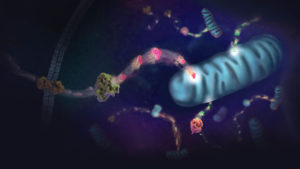
Metabolism underpins numerous cellular processes. Without it, cells would not grow, divide, synthesize or secrete. Another pathway, autophagy, degrades unwanted cellular materials, helping to maintain cell health. With these opposing roles, is there a connection between autophagy and metabolism? As it turns out, the answer is yes. Because molecules degraded by autophagy are recycled and fed into metabolism pathways as precursor compounds. There are interesting implications as a result of this connection, ones that affect cancer cells as described in a recent Cell Metabolism review article.

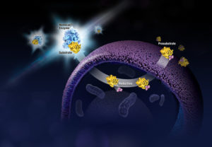
Autophagic flux, the process by which molecules and organelles are directed to the autophagosome, fuse with the lysosome and are degraded, involves a selective process that determines the cargo carried within the autophagosome. Autophagy-related genes (ATGs) direct the process and particular receptor proteins bind the cargo. What is interesting about the connection among cancer, autophagy and metabolism is the complexity of the role that autophagy plays in cancer. While autophagy was thought to act in a more tumor suppressive manner as shown when one copy of an ATG6 analogous gene in mice was deleted and the other left unaltered, and malignant tumors developed, but in mice mosaic for ATG5 deletions, the inhibition of autophagy resulted in benign tumors in the liver. This latter experiment suggested autophagy was needed for cancer progression, a hypothesis reinforced by the lack of ATG mutations in human cancers.

Continue reading “How Autophagy Feeds Cancer’s Need for Metabolites”

Finding Chinks in the Armor: Cancer’s Need for Metabolites

Illustration of energy metablism in cell.

Cancer has been studied for decades by scientists trying to find a vulnerability to exploit and testing compounds to develop as potential drugs. As the “Emperor of All Maladies”, cancer has proven itself to be a wily beast with many varieties of genetic mutations for eluding cellular control, tireless in its ability to divide and spread. In the end, a cancer cell is still a cell and subject to its environment even though cancer does not play by the same rules as the normal cells that exist around it. To be able to grow, a cell needs access to metabolites, molecules needed for building the materials and machinery needed by the cell to function and divide. These requirements also offer potential pathways to target for halting cancer growth and spread.

All cells use glucose to generate ATP, but normal and cancer cells differ in how glucose is converted to ATP. Most cells use glucose in oxidative phosphorylation, but cancer cells use aerobic glycolysis, converting glucose to lactate without oxygen. This Warburg effect (glucose converted to lactate) is a hallmark of cancer cells as they take up glucose at a much higher rate than normal cells. Blocking glucose uptake is one way to target cancer cells. While 2-deoxyglucose (2DG) has been shown to slow glucose uptake in vitro, the compound proved toxic in clinical trials and lower dosages do not seem to be an effective treatment against cancer. While not an ideal drug target, glucose uptake has been helpful in monitoring cancer response to therapies via fluorodeoxyglucose positron emission tomography (FDG-PET).

Continue reading “Finding Chinks in the Armor: Cancer’s Need for Metabolites”

Genomic Breakthroughs One Letter at a Time

Breakthrough at the human genome project

In honor of Human Genome Month, I delved into our Cartoon Lab archives to retrieve this example of the excitement that occurred while sequencing the Human Genome Project.

For more entertaining science cartoons, visit our Cartoon Lab.

Celebrating Women in Science

By US Environmental Protection Agency [Public domain], via Wikimedia Commons

February 11 is the International Day of Women and Girls in Science, a reminder that there is still a gender gap in science. Despite the obstacles that women need to overcome, their contributions to field of science have benefited not only their fellow researchers but also their fellow humans. From treatments for diseases to new discoveries that opened up entire fields, women have advanced knowledge across the spectrum of science. Below is a sampling of the achievements of just a few women in science. What other living female scientist or inventor might you add?

Hate malaria? You can thank Tu Youyou for discovering artemisinin and dihydroartemisinin, compounds that are used to treat the tropical disease and save numerous lives. Her discovery was so significant, she received the 2015 Nobel Prize in Physiology or Medicine.

Continue reading “Celebrating Women in Science”

Two Epigenetic Targets Are More Effective Than One

Lysine-specific histone demethylase 1 (LSD1) via Wikimedia Commons

Epigenetics is a new and exciting territory to explore as we understand more about the role it plays in gene silencing and expression. Because epigenetic regulation of gene expression is caused by specific modification of histone proteins (e.g., methylation) that play a role in disease states like cancer, enzymes like histone deacetylases (HDACs) become viable drug targets. One drawback to inhibiting proteins that modify histones is even when selectively targeting HDACs, the effects can be far ranging with multiple HDAC-containing protein complexes found throughout the cell. These broad effects minimize the effectiveness of an inhibitor, caught between efficacy and toxicity. A recent article in Nature Communications explored how using a single compound to target two epigenetic enzymes was more effective than any individual inhibitor or combination of inhibitors. Continue reading “Two Epigenetic Targets Are More Effective Than One”

Hot Wings and Snow Birds: A Study of Genetic Selection in Chickens

African chicken breed Boschvelder. Image copyright ICBH GROUP.

This past summer, I visited the county fair and stopped by the animal barn to look at some of the poultry on display. Specifically, I wanted to see examples of the breeds of chickens available that I may be interested in adding to my flock. Rather than each chicken in their display cage being labeled with a bird’s breed, each cage listed the geographic origin of the chicken within such as Asiatic, Continental or American. This did not benefit my search for potential new members of my flock, but intrigued me enough that I wanted to find out how my flock of 19 hens and pullets would be characterized. Using the classes delineated by the Wisconsin State Fair, my feathered ladies break down to 12 American, 4 English and 3 Continental chickens. There are also classes for Mediterranean and Asiatic (and Other). I live in a part of the United States that gets cold, snowy weather for what seems like six months out of the year, weather that my chickens seem to take in stride. But in other places in the world, heat is the name of the game for the poultry strutting there. In a Genes, Genomics, Genetics publication, Fleming et al. wanted to know if there were genetic differences in Northern European and African chickens that might be caused by their environment.

Continue reading “Hot Wings and Snow Birds: A Study of Genetic Selection in Chickens”

Reveal More Biology: How Real-Time Kinetic Cell Health Assays Prove Their Worth

What if you could uncover a small but significant cellular response as your population of cells move toward apoptosis or necrosis? What if you could view the full picture of cellular changes rather than a single snapshot at one point? You can! There are real-time assays that can look at the kinetics of changes in cell viability, apoptosis, necrosis and cytotoxicity—all in a plate-based format. Seeking more information? Multiplex a real-time assay with endpoint analysis. From molecular profiling to complementary assays (e.g., an endpoint cell viability assay paired with a real-time apoptosis assay), you can discover more information hidden in the same cells during the same experiment.

Whether your research involves screening a panel of compounds or perturbing a regulatory pathway, a more complete picture of cellular changes gives you the benefit of more data points for better decision making. Rather than assessing the results of your experiment using a single time point, such as 48 hours, you could monitor cellular changes at regular intervals. For instance, a nonlytic live-cell reagent can be added to cultured cells and measurements taken repeatedly over time. Pairing a real-time cell health reagent with a detection instrument that can maintain the cells at the correct temperature means you can automate the measurements. These repeated measurements over time reveal the kinetic changes in the cells you are testing, giving a real-time status update of the cellular changes from the beginning to the end of your experiment. Continue reading “Reveal More Biology: How Real-Time Kinetic Cell Health Assays Prove Their Worth”

Six (and a Half) Reasons to Quantitate Your DNA

Knowing how much DNA you have is fundamental to successful experiments. Without a firm number in which you are confident, the DNA input for subsequent experiments can lead you astray. Below are six reasons why you should quantitate your DNA.

6. Saving time by knowing what you have rather than repeating experiments. If you don’t quantitate your DNA, how certain can you be that the same amount of DNA is consistently added? Always using the same volume for every experiment does not guarantee the same DNA amount goes into the assay. Each time there is a new purified DNA sample, the chances that you have the same quantity as before are lessened. Consequently, without knowing the DNA concentration of the sample you are using, the amount of input DNA cannot be guaranteed and experiments may have to be repeated.

Continue reading “Six (and a Half) Reasons to Quantitate Your DNA”

Revealing Time of Death: The Microbiome Edition

Forensic analysts have long sought precision when determining time of death. While on crime scene investigation television shows, the presence of insects always seems to reveal when a person died, there are many elements to account for, and the probable date may still not be accurate. Insects arrive days after death if at all (e.g., if the body is found indoors or after burial), and the stage of insect activity is influenced by temperature, weather conditions, seasonal variation, geographic location and other factors. All this makes it difficult to estimate the postmortem interval (PMI) of a body discovered an unknown time after death. One way to make estimating PMI less subjective would be to have calibrated molecular markers that are easy to sample and are not altered by environmental variabilities.

Bacterial communities called microbiomes have been frequently in the news. The influence of these microbes encompass living creatures and the environment. Not surprisingly, research has focused on the influence of microbiomes on humans. For example, changes in gut microbiome seem to affect human health. Intriguingly, microbiomes may also be a key to determining time of death. The National Institute of Justice (NIJ) has funded several projects focused on the forensic applications of microbiomes. One focus involves the necrobiome, the community of organisms found on or around decomposing remains. These microbes could be used as an indicator of PMI when investigating human remains. Recent research published in PLOS ONE examined the bacterial communities found in human ears and noses after death and how they changed over time. The researchers were interested in developing an algorithm using the data they collected to estimate of time of death.

Continue reading “Revealing Time of Death: The Microbiome Edition”

Preventing Viral Infection by Blocking Cellular Receptors with a Tethered Antibody

Finding a way to neutralize or block infection by HIV has long been pursued by viral researchers. Various treatments have been developed, driven by the need to find effective drugs to manage HIV in infected individuals. The ultimate goal is to develop a vaccine to prevent HIV from even taking hold in the body. With all of our knowledge about the cellular receptors HIV needs to enter the cell, there has to be a method to prevent a viral particle from binding and being internalized. Many researchers are pursuing neutralizing antibodies to the virus as one method. What about antibodies that target the cellular receptor recognized by the virus? An article published in Proceedings of the National Academy of Sciences, antibodies to cellular receptors for rhinovirus and HIV were tethered to the plasma membrane and tested for the ability to prevent infection.

Continue reading “Preventing Viral Infection by Blocking Cellular Receptors with a Tethered Antibody”