3D Cell Culture Models: Challenges for Cell-Based Assays

3D Cell Culture Spheroid

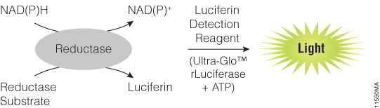
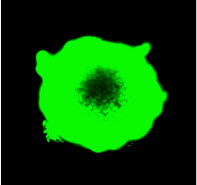
In 3D cell culture models, cells are grown under conditions that allow the formation of multicellular spheroids or microtissues. Instead of growing in a monolayer on a plate surface, cells in 3D culture grow within a support matrix that allows them to interact with each other, forming cell:cell connections and creating an environment that mimics the situation in the body more closely than traditional 2D systems. Although 3D cultures are designed to offer a more physiologically accurate environment, the added complexity of that environment can also present challenges to experimental design when performing cell-based assays. For example, it can be a challenge for assay reagents to penetrate to the center of larger microtissues and for lytic assays to disrupt all cells within the 3D system.

Earlier this week Terry Riss, a Senior Product Specialist at Promega, presented a Webinar on the challenges of performing cell-based assays on microtissues in 3D cell culture. During the Webinar, Terry gave an overview of the different methods available for 3D cell culture, providing a description of the advantages of each. He then discussed considerations for designing and optimizing cell-based  assays for use in 3D culture systems, providing several  recommendations to keep in mind when performing cell viability assays on larger microtissue samples.

Continue reading “3D Cell Culture Models: Challenges for Cell-Based Assays”

Measuring Changing Metabolism in Cancer Cells

Because of the central role of energy metabolism in health and disease, and its effect on other cellular processes, assays to monitor changes in cellular metabolic state have wide application in both basic research and drug discovery. In the webinar “Tools for Cell Metabolism: Bioluminescent NAD(P)/NAD(P)H-Glo™ Assays” Jolanta Vidurigiene, a Senior Research Scientist at Promega, introduces three metabolism assays for measuring oxidized and reduced forms of NAD and NADP.

In this webinar, Jolanta provides background information on why it is important to be able to accurately measure metabolites such as NAD/NADH and NADP/NADPH. She outlines the roles of each, and highlights some of the challenges involved in developing assays that can accurately measure these metabolites. She discusses key considerations for successful NAD(P)/NAD(P)H assays and provides examples of how to use these assays to measure either total (both oxidized and reduced) forms of NAD and NADP, or to measure oxidized and reduced forms individually in a single assay plate.

NAD(P)H-Glo™ Assay Mechanism

Continue reading “Measuring Changing Metabolism in Cancer Cells”

A Quick Method for A Tailing PCR Products

PCR experiment and products, pipette tip, tube in researcher's hand.
PCR is a common technique used in research labs to amplify DNA.

Some thermostable DNA polymerases, including Taq, add a single nucleotide base extension to the 3′ end of amplified DNA fragments. These polymerases usually add an adenine, leaving an “A” overhang. There are several approaches to overcome the cloning difficulties presented by the presence of A overhangs on PCR products. One method involves treating the product with Klenow to create a blunt-ended fragment for subcloning. Another choice is to add restriction sites to the ends of your PCR fragments. You can do this by incorporating the desired restriction sites into the PCR primers. After amplification, the PCR product is digested and subcloned into the cloning vector. Take care when using this method, as not all restriction enzymes efficiently cleave at the ends of DNA fragments, and you may not be able to use every restriction enzyme you desire. There is some useful information about cutting with restriction sites close to the end of linear fragments in the Restriction Enzyme Resource Guide. Also, some restriction enzymes require extra bases outside the recognition site, adding further expense to the PCR primers as well as risk of priming to unrelated sequences in the genome.

Continue reading “A Quick Method for A Tailing PCR Products”

Bacteria and Viruses as Cancer Treatments

Over a hundred years ago William B Coley, the “Father of Immunotherapy”, discovered that injection of bacteria or bacterial toxins into tumors could cause those tumors to shrink. The introduction of bacteria had the side-effect of stimulating the immune system to attack the tumor. The field of cancer immunotherapy research—which today includes many different approaches for generating anti-tumor immune responses—originated with these early experiments.

Use of bacteria is one way to stimulate the immune system to attack cancer cells, others include use of cytokines, immune checkpoint blockades and vaccines. This Nature animation provides a simple overview of these methods.

Continue reading “Bacteria and Viruses as Cancer Treatments”

How to Take Care of Your Pipettes

Last updated: 1/22/21

what not to do with your pipettes

Pipettes are such a routine part of everyday life in the lab that it can be easy to take them for granted.  Their accuracy is vital, and there are many things we can adopt as best practices for success. Here are a few tips (no pun intended) gathered from around the Web by Kim Steinhauser of the Promega Metrology Department–the group charged with keeping our pipettes and other lab equipment functional and accurate.

Continue reading “How to Take Care of Your Pipettes”

The Pan-Cancer Atlas: “The End of the Beginning”

In April 2018, a series of 27 papers representing the most comprehensive genomic analysis of human cancers to date was published in Cell Press journals.

The collection constitutes the final outputs from the Cancer Genome Atlas (TCGA) project, a collaboration between the National Cancer Institute (NCI) and the National Human Genome Research Institute (NHGRI) involving analysis of over 11,000 tumors representing 33 different cancers. The many research teams involved analyzed tumor DNA, mRNA, miRNA and chromatin, comparing them to matched normal cellular genomes to perform a complete molecular characterization of cancer-specific changes. The results have been presented with much hope that open access to this type of comprehensive analysis will build on recent advances in understanding tumor biology and spur further progress in developing new approaches to treatment. (See this news item for more detail).

The Pan-Cancer Atlas results are collected on a cell.com portal, where they are presented in three collections grouped by topic: Cell of Origin, Oncogenic Processes and Signaling Pathways. Each collection is accompanied by a “Flagship” paper introducing the topic and summarizing the findings. It seems fitting that these findings have been published in #HumanGenomeMonth. This comprehensive analysis of the genomic and metagenomic profiles of tumors illustrates one powerful application of the type of genomic analysis pioneered by the original Human Genome Project, and shows just how much has been made possible since the initial publication of the human genome fifteen years ago.

Continue reading “The Pan-Cancer Atlas: “The End of the Beginning””

A Better Way to Understand How and Why Cells Die

Real-time, up-to-the-minute access to information provides new opportunities for scientists to monitor cellular events in ever more meaningful ways. Real-time cytotoxicity and cell viability assay reagents now allow constant monitoring of cell health status without the need to lyse or remove aliquots from plates for measurement. With a real-time approach, data can be collected from cell cultures or microtissues at multiple time points after addition of a drug compound or other event, and the response to treatment continually observed.

The CellTox™ Green assay is a real-time assay that monitors cytotoxicity using a fluorescent DNA binding dye, which binds DNA released from cells upon loss of membrane integrity. The dye cannot enter intact, live cells and so fluorescence only occurs upon cell death, correlating with cytotoxicity. Here’s a quick overview showing how the assay works:

More Data Using Fewer Samples and Reagents
The ability to continually monitor cytotoxicity in this way makes it easy to conduct more than one type of analysis on a single sample. Assays can be combined to determine not only the timing of cytotoxicity, but to also understand related events happening in the same cell population. As long as the readouts can be distinguished from one another multiple assays can be performed in the same well, providing more informative data while using less cells, plates and reagents.

Combining assays in this way can reveal critical information regarding mechanism of cell death. For example, assay combinations can be used to determine whether cells are dying from apoptosis or necrosis, or to distinguish nonproliferation from cell death. Combining CellTox Green with an endpoint luminescent caspase assay or a real-time apoptosis assay allows you to determine whether observed cytotoxic effects are due to apoptosis. Cytotoxic and anti-proliferative effects can be distinguished by combining the cytotoxicity assay with a luminescent or fluorescent cell viability assay.

Continue reading “A Better Way to Understand How and Why Cells Die”

A Cell Viability Assay for Today

Valued for ease of use and scalability, plate-based, bioluminescent cell viability assays are widely used to support research in biologics, oncology and drug discovery.

Cell viability assays are a bread-and-butter method for many researchers using cultured cells —everyday lab tools that are a part of many newsworthy papers, but rarely make news themselves.

Over time, cell viability assays have become easier to use and more “plug ‘n play”. Among modern assays, luminescent plate-reader based systems have been a favorite for several years because of their superior sensitivity, robustness, simple protocols and uncomplicated equipment requirements (all you need is a plate-reading luminometer). These qualities combine to allow easy scalability and adaptability from bench research to high throughput applications.

CellTiter-Glo® Luminescent Cell Viability Assay is an accepted go-to viability assay for many researchers. The assay measures ATP as an indicator of metabolically active cells. A quick search on Google Scholar returns 3,990 CellTiter-Glo results for 2017 and over 500 so far in January and February of 2018. A sampling of these recent publications gives a snapshot of some of the ways the CellTiter-Glo assay is used to support key areas of research today.

Does a treatment kill cells?

The obvious application of a cell viability assay is to understand whether cells are alive. In cancer research, the CellTiter-Glo assay is often used to confirm killing of tumor cells and to verify that normal cells survive. Therefore, these assays are a key part of the evaluation and screening of drug candidates and other therapies for cancer. Many papers reporting use of CellTiter-Glo are developing and evaluating the effectiveness of novel anti-cancer treatments. Continue reading “A Cell Viability Assay for Today”

The Bacteria that are Good for Us

Chains of Streptococci

Salmonella. Streptococcus. Shigella. The most well-known bacteria are those that cause disease. Our relationship with them is one of combat. With good reason, we look for ways to avoid encountering them and to eliminate them when we do meet.

But not all bacteria are bad for us. Of course we have known for years that we are colonized by harmless bacteria, but recently, studies on the human microbiome have revealed many surprising things about these bacterial tenants. Studies are showing that the teeming multitudes of organisms living in and on the human body are not just harmless bystanders, but complex, interrelated communities that can have profound effects on our health.

Three studies published in Science in 2018 add more to the growing body of microbiome surprises, showing that certain gut bacteria are not only good for us, but may even be required for the effectiveness of some anti-cancer immunotherapies.

Continue reading “The Bacteria that are Good for Us”

Five Ways to Explain the CRISPR Story Without Delivering a Lecture

Recently a FaceBook friend of mine (who is not a scientist) shared a video from WIRED Science where the concept of CRISPR is explained at 5 Levels of Difficulty— for a 7 year old, a teenager, a college student, a grad student and a CRISPR expert.

First it was pretty amazing to me that my non-scientist friends are interested enough in learning about CRISPR to share this type of information—perhaps showing just how popular and exciting the method has become. People outside the scientific field are hearing a lot about it, and are curious to know more.

This video does a great job of explaining the technique for all its intended audiences. It also is a nice illustration of how to share information in an easily understandable format. Even with the 7 year old and 14 year old, the information is shared in a conversational way, with everyone involved contributing to sharing information about CRISPR.

Really nice. Here’s the WIRED video:


Continue reading “Five Ways to Explain the CRISPR Story Without Delivering a Lecture”