How Prostate Cancer Cells Survive Glucose Deprivation

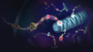
Illustration of energy metablism in cell.

Glucose is an energy metabolite necessary for cellular survival and growth whether or not the cell is part of a tumor. Not only do cancer cells switch from oxidative phosphorylation to aerobic glycolysis (the Warburg effect) to gain more glucose, a hallmark of cancer, but they also increase the amount of glucose taken up from the surrounding extracellular space. However, the lack of glucose can have a negative effect on cells, causing them to become apoptotic in the absence of this metabolite. Cancer cells have methods to get around the requirement for glucose, including upregulating glucose transporters to improve access to the energy metabolite. In this Redox Biology article, researchers describe how activating androgen receptor in response to a lack of glucose affects the amount of GLUT1 expressed on prostate cancer cells, making the cells resistant to glucose deprivation.

To set the stage, two prostate cancer cell lines, LNCaP, an androgen-sensitive cell line, and LNCaP-R, an androgen-insensitive cell line, were deprived of glucose. Both cell lines showed signs of cell death, but LNCaP-R cells died in greater numbers. To probe how LNCaP cells died, several inhibitors (a pan-caspase inhibitor, two necroptosis inhibitors and a ferroptosis inhibitor) were added but did not change the way the cells died. However, an autophagy inhibitor enhanced cell death, suggesting the cells were necrotic not apoptotic. Teasing apart if the necrosis of LNCaP cells was due to glucose availability or merely disrupted glycolysis, the glucose analog 2DG was added to the medium with glucose. The cells survived when treated with 2DG, suggesting it was the absence of glucose that induced necrosis. When LNCaP cells were cultivated in medium that replaced glucose with mannose or fructose, the cells survived, another point in favor of sugar depletion causing cell death.

Continue reading “How Prostate Cancer Cells Survive Glucose Deprivation”
How Autophagy Feeds Cancer’s Need for Metabolites

Illustration of energy metablism in cell.

Metabolism underpins numerous cellular processes. Without it, cells would not grow, divide, synthesize or secrete. Another pathway, autophagy, degrades unwanted cellular materials, helping to maintain cell health. With these opposing roles, is there a connection between autophagy and metabolism? As it turns out, the answer is yes. Because molecules degraded by autophagy are recycled and fed into metabolism pathways as precursor compounds. There are interesting implications as a result of this connection, ones that affect cancer cells as described in a recent Cell Metabolism review article.

Autophagic flux, the process by which molecules and organelles are directed to the autophagosome, fuse with the lysosome and are degraded, involves a selective process that determines the cargo carried within the autophagosome. Autophagy-related genes (ATGs) direct the process and particular receptor proteins bind the cargo. What is interesting about the connection among cancer, autophagy and metabolism is the complexity of the role that autophagy plays in cancer. While autophagy was thought to act in a more tumor suppressive manner as shown when one copy of an ATG6 analogous gene in mice was deleted and the other left unaltered, and malignant tumors developed, but in mice mosaic for ATG5 deletions, the inhibition of autophagy resulted in benign tumors in the liver. This latter experiment suggested autophagy was needed for cancer progression, a hypothesis reinforced by the lack of ATG mutations in human cancers.

Continue reading “How Autophagy Feeds Cancer’s Need for Metabolites”

A “Spare Tire” for Proto-oncogene Promoters

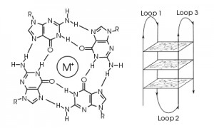
A guanine tetrad (left) and G-quadruplex (right). Image courtesy of Wikimedia Commons.

Proto-oncogenes are genes that organisms rely on for normal growth and development but, when mutated or dysregulated, can cause cells to grow uncontrollably, resulting in cancer and metastasis. In some cases, a single DNA mutation is sufficient for cancer to develop. Why then, do so many proto-oncogene promoters contain strings of guanine residues, which are extremely vulnerable to DNA damage from factors such as oxidative stress and hyperinflammation, to control transcription levels? From an evolutionary viewpoint, this is a contradiction: DNA sequences that are the most vulnerable to damage and mutation are key to regulating one of the cell’s most dangerous classes of genes. This seems to be a recipe for genomic instability and disease. Fortunately, evolution has provided a very clever solution to this potential problem.

Continue reading “A “Spare Tire” for Proto-oncogene Promoters”

Piecing the Puzzle Together: Using Multiple Assays to Better Understand What Is Happening with Your Cells

You often need several pieces of information to really understand what is happening within a cell or population of cells. If your cells are not proliferating, are they dying? Or, are you seeing cytostasis? If they are dying, what is the mechanism? Is it apoptosis or necrosis? If you are seeing apoptosis, what is the pathway: intrinsic or extrinsic?

If you are measuring expression of a reporter gene and you see a decrease in expression, is that decrease due to transfection inefficiencies, cytotoxicity, or true down regulation of your reporter gene?

To investigate these multiple parameters, you can run assays in parallel, but that requires more sample, and sample isn’t always abundant.

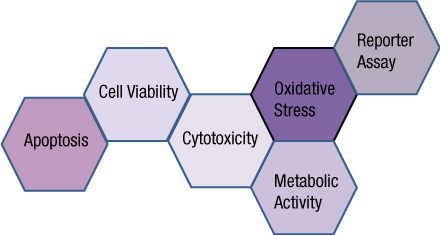
Multiplexing assays allows you to obtain information about multiple parameters or events (e.g., reporter gene expression and cell viability; caspase-3 activity and cell viability) from a single sample. Multiplexing saves sample, saves time and gives you a more complete picture of the biology that is happening with your experimental sample.

What information do you need about your cells to complete the picture?

Multiplexing assay reagents to measure biomarkers in the same sample has often been considered an application only accomplished with antibodies or dyes and sophisticated detection instrumentation. However, Promega has developed microwell plate based assays for cells in culture that allow multiplexed detection of biomarkers in the same sample well using standard multimode multiwell plate readers. Continue reading “Piecing the Puzzle Together: Using Multiple Assays to Better Understand What Is Happening with Your Cells”