
A Cell Viability Assay for Today

Valued for ease of use and scalability, plate-based, bioluminescent cell viability assays are widely used to support research in biologics, oncology and drug discovery.

Cell viability assays are a bread-and-butter method for many researchers using cultured cells —everyday lab tools that are a part of many newsworthy papers, but rarely make news themselves.

Over time, cell viability assays have become easier to use and more “plug ‘n play”. Among modern assays, luminescent plate-reader based systems have been a favorite for several years because of their superior sensitivity, robustness, simple protocols and uncomplicated equipment requirements (all you need is a plate-reading luminometer). These qualities combine to allow easy scalability and adaptability from bench research to high throughput applications.

CellTiter-Glo® Luminescent Cell Viability Assay is an accepted go-to viability assay for many researchers. The assay measures ATP as an indicator of metabolically active cells. A quick search on Google Scholar returns 3,990 CellTiter-Glo results for 2017 and over 500 so far in January and February of 2018. A sampling of these recent publications gives a snapshot of some of the ways the CellTiter-Glo assay is used to support key areas of research today.

Does a treatment kill cells?

The obvious application of a cell viability assay is to understand whether cells are alive. In cancer research, the CellTiter-Glo assay is often used to confirm killing of tumor cells and to verify that normal cells survive. Therefore, these assays are a key part of the evaluation and screening of drug candidates and other therapies for cancer. Many papers reporting use of CellTiter-Glo are developing and evaluating the effectiveness of novel anti-cancer treatments. Continue reading “A Cell Viability Assay for Today”
Piecing the Puzzle Together: Using Multiple Assays to Better Understand What Is Happening with Your Cells

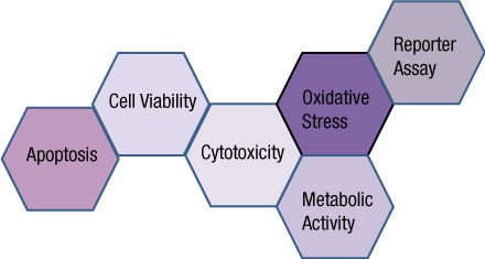
You often need several pieces of information to really understand what is happening within a cell or population of cells. If your cells are not proliferating, are they dying? Or, are you seeing cytostasis? If they are dying, what is the mechanism? Is it apoptosis or necrosis? If you are seeing apoptosis, what is the pathway: intrinsic or extrinsic?

If you are measuring expression of a reporter gene and you see a decrease in expression, is that decrease due to transfection inefficiencies, cytotoxicity, or true down regulation of your reporter gene?

To investigate these multiple parameters, you can run assays in parallel, but that requires more sample, and sample isn’t always abundant.

Multiplexing assays allows you to obtain information about multiple parameters or events (e.g., reporter gene expression and cell viability; caspase-3 activity and cell viability) from a single sample. Multiplexing saves sample, saves time and gives you a more complete picture of the biology that is happening with your experimental sample.

What information do you need about your cells to complete the picture?

Multiplexing assay reagents to measure biomarkers in the same sample has often been considered an application only accomplished with antibodies or dyes and sophisticated detection instrumentation. However, Promega has developed microwell plate based assays for cells in culture that allow multiplexed detection of biomarkers in the same sample well using standard multimode multiwell plate readers. Continue reading “Piecing the Puzzle Together: Using Multiple Assays to Better Understand What Is Happening with Your Cells”