Looking Back: Cell-Free Expression Systems Helped to Characterize Proteins Involved in Hypoxia Response

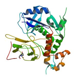
Structur of a HIF-1a-pVHL-ElonginB-ElonginC complex
Structure of a HIF-1a-pVHL-ElonginB-ElonginC complex

William G. Kaelin Jr., Sir Peter J. Ratcliffe and Gregg L. Semenza were awarded the 2019 Nobel Prize in Physiology or Medicine for their discoveries of how cells sense and adapt to oxygen availability.

Kaelin and Ratcliffe’s labs focused their efforts on the transcription factor HIF (hypoxia-inducible factor). This transcription factor is critical in the cellular adaptation of to changes in oxygen availability.

When oxygen levels are elevated cells contain very little HIF. Ubiquitin is added to the HIF protein via the VHL complex and it is degraded in the proteasome.  When oxygen levels are low (hypoxia) the amount of HIF increases.

In 2001 both groups published articles characterizing the interaction between VHL and HIF, and these articles were referenced by the Nobel Prize Organization in their press release about this year’s award. (1,2). Both studies demonstrated that under the normal oxygen conditions hydroxylation of proline residue P564 enabled VHL to recognize and bind to HIF.

The use of cell free expression (i.e., TNT Coupled Transcription/Translation System) by both labs was key in the characterization of the VHL:HIF interaction The labs utilized HIF and VHL 35-S labeled proteins generated via the TNT system under both normal or in a hypoxic work station to:

  • Determine the affect of ferrous chloride and cobaltous chloride on the interaction
  • Map the specific region of HIF required for the interaction to occur (556-574)
  • Determine the effect of HIF point mutations on the interaction
  • Use synthetic peptides to block the interaction
  • Conclude that a factor in mammalian cells was necessary for the interaction to occur.

Literature Cited

  1. Ivan, M et al. (2001) HIF Targeted for VHL-Mediated Destruction by Proline Hydroxylation: Implications for O2 Sensing. Science 292: 464–67.
  2. Jaakkola, P. et al. (2001) Targeting of HIF-α to the von Hippel-Lindau Ubiquitylation of Complex by O2– Regulated Prolyl Hydroxylation. Science 202, 468–72 .
Related Posts

Cell Free Application: Characterization of Long Non-coding RNA Inhibition of Transcription

Long noncoding RNAs have been shown to regulate chromatin states, transcriptional activity and post transcriptional activity (1). Only a few studies have observed long non-coding RNAs modulating the translational process (2). The noncoding RNA BC200 has been shown to inhibit translation by interacting with the translation initiation factors, eIF4A and eIF4B.

To characterize how BC200 translational inhibition could be controlled,  a variety of RNAs were transcribed/translated in vitro using the TNT system (Cat. #L4610) from Promega. To each transcription/translation reaction, BC900 RNA, hnRNPE1 and hnRNE2 proteins were added. Inhibition of BC200 activity was noted when proteins were successful expressed (3).

Literature Cited

  1.  Sosinska, P et.al. (2015) Intraperitoneal invasiveness of ovarian cancer from the cellular and molecular perspective. Ginekol. Pol. 86, 782–86.
  2. Geisler, S. and Coller, J. (2013) RNA in unexpected places: long non-coding RNA functions in diverse cellular contexts. Nat.Rev. Mol. Cell. Bio. 14,699–12.
  3. Jang, S. et. al. (2017) Regulation of BC200 RNA-mediated translation inhibition by hnRNP E1 and E2. FEBS Letters. 591, 393–5.

Protease K Protection Assay: Cell Free Expression Application

Microsomal vesicles are used to study cotranslational and initial posttranslational processing of proteins. Processing events such as signal peptide cleavage, membrane insertion, translocation and core glycosylation can be examined by the transcription/translation of the appropriate DNA in the TNT® Lysate Systems when used with microsomal membranes.

The most general assay for translocation makes use of the protection afforded the translocated domain by the lipid bilayer of the microsomal membrane. In this assay protein domains are judged to be translocated if they are observed to be protected from exogenously added protease. To confirm that protection is due to the lipid bilayer addition of 0.1% non-ionic detergent (such as Triton® X-100) solubilizes the membrane and restores susceptibility to the protease.

Are you looking for proteases to use in your research?
Explore our portfolio of proteases today.

Many proteases have proven useful for monitoring translocation in this fashion including Protease K or Trypsin.

The following are examples illustrating this application:

  1. Minn, I. et al. (2009) SUN-1 and ZYG-12, mediators of centrosome-nucleus attachment, are a functional SUN/KASH pair in Caenorhabditis elegans. Mol. Biol. Cell. 20, 4586–95.
  2. Padhan, K. et al. (2007) Severe acute respiratory syndrome coronavirus Orf3a protein interacts with caveolin. J.Gen.Virol. 88, 3067–77.
  3. Tews, B.A. et al. (2007) The pestivirus glycoprotein Erns is anchored in plane in the membrane via an amphipathic helix. J.Biol.Chem. 282, 32730–41.
  4. Pidasheva, S. et al. (2005) Impaired cotranslational processing of the calcium-sensing receptor due to signal peptide missense mutations in familial hypocalciuric hypercalcemia. Hum. Mol. Gen. 14, 1679–90.
  5. Smith, D. et al. (2002) Exogenous peptides delivered by ricin require processing by signal peptidase for transporter associated with antigen processing-independent MHC class I-restricted presentation. J. Immun. 169, 99–107.