Galloping to Greatness: Meet Kurt the First Cloned Przewalski’s Horse

On August 6, 2020, the first successfully cloned Przewalski’s horse was born at the Texas-based veterinary facility, Timber Creek Veterinary, along with a new hope for restoring some much-needed genetic diversity to the species. The successful birth of this foal is the culmination of the collaborative efforts between Revive & Restore, San Diego Zoo Global (SDZG), and ViaGen Equine, and lays the groundwork as an important model for future conservation efforts.

Kurt the first cloned Przewalsk'si horse
Kurt at Timber Creek Veterinary, 09/28/20.
Photo by Scott Stine.

The new Przewalski’s foal (pronounced “shuh-VAL-skees”) has been affectionately dubbed Kurt, in honor of noted animal conservationist, geneticist and pathologist, Dr. Kurt Benirschke. Dr. Benirschke played an instrumental role in founding the Frozen Zoo®, a genetic library comprised of cryopreserved cell lines of endangered species. Established in the 1970s, this collection was built on a foundation of prescient hope, banking on the future development of reproductive and cloning technologies that did not yet exist.

Now thanks to his foresight, that gamble is paying off and the fruits of that labor are literally being brought to life almost 50 years later through Kurt the foal, who is as adorable as he is important to the future of his kind.

Continue reading “Galloping to Greatness: Meet Kurt the First Cloned Przewalski’s Horse”

XpressAmp™ Direct Amplification: Simplified and Accelerated Time to qPCR Results

As the SARS‐CoV‐2 pandemic continues to rage across the United States and around the globe, the demand for COVID‐19 testing is increasing. The vast majority of the COVID-19 assays use RT‐qPCR to detect the viral RNA in patient samples such as nasopharyngeal swabs, which are collected and stored in viral or universal transport media (VTM/UTM). The general workflow for these COVID‐19 assays can be broken down as follows:

  1. Collect and store patient samples
  2. Ship samples to testing laboratory
  3. Extract RNA from samples
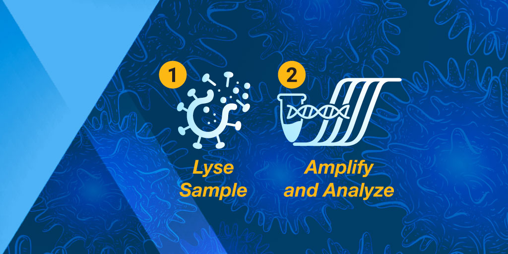
  4. Amplify and analyze samples

While many companies who manufacture the products that are used in these steps have been able to adapt and significantly increase their production capacities, there are still gaps between the supply of these products and the global test demand. Both the sample collection and storage step and the RNA extraction/purification step have a tendency to bottleneck and experience supply constraints. One way to address these bottlenecks and expand production capacity for these in‐demand products is to evaluate the viability of skipping a step in the workflow, without hindering the ability to detect viral RNA from samples.

Continue reading “XpressAmp™ Direct Amplification: Simplified and Accelerated Time to qPCR Results”

A Specific and Sensitive Matter: The Trouble With COVID-19 Antibody Tests

From the beginning of this pandemic, scientists around the world have been working around the clock in pursuit of answers that can effectively combat the SARS-CoV-2 virus. One of hardest things for people to grapple with, is all the unknowns: When will this end? When can I safely visit my friends and family again? What if I have it or had it and I don’t even know it?  

The increased availability of serological testing has helped ease people’s minds about their personal COVID-19 status. From a distance, serological testing may seem like a huge milestone in the marathon that is this pandemic. Unfortunately, many of these tests provide murky and inconsistent results.

Continue reading “A Specific and Sensitive Matter: The Trouble With COVID-19 Antibody Tests”

From Live Cells to Lysates: Adapting NanoBiT to a Biochemical Assay Format

The ability to target protein interactions with low solubility or weak binding affinities can present a significant challenge when it comes to drug screening. The beauty of these types of challenges we often face in the lab is that finding solutions to these problems doesn’t necessarily require brand new tools. Sometimes we already have the right tools in our arsenal and, with just a little creativity and collaboration, they can be adapted to address the challenge at hand.

In the following video, Dr. Mohamed (Soly) Ismail, a Postdoctoral Fellow at the Downward Lab of the Francis Crick Institute, presents the perfect example of this with his novel approach to the NanoBiT® Protein:Protein Interaction Assay. Through a collaboration with Promega R&D Scientists, Dr. Ismail has translated the assay into a cell-free, biochemical format, termed the NanoBiT Biochemical Assay (NBBA).

Watch the subtitled version of the video here >

Continue reading “From Live Cells to Lysates: Adapting NanoBiT to a Biochemical Assay Format”

Out-FOXOing High-Stage Neuroblastoma

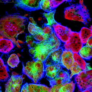
Fluorescence microscopy of neuroblastoma cells.

In recent years, scientists have been hot on the trail of transcription factor FOXO3, tracing its involvement in various tumor-centric activities comprising the many trademarks of cancer, from drug resistance to metastasis to tumor angiogenesis.

FOXO3 is a member of the O sub-class of the forkhead box family of transcription factors. The forkhead box (FOX) family is characterized by a fork head DNA-binding domain (DBD), comprised of around 100 amino acids. They have also proven themselves to be a family of many hats, functioning in diverse roles ranging from metabolism, immunology, cell-cycle control, development, as well as cancer (1). The forkhead box O (FOXO) sub-class alone has demonstrated involvement in a variety of cellular outcomes, from drug resistance and longevity to apoptosis induction.

Due to its pro-apoptotic and anti-proliferative proclivity, FOXO3 has been previously identified as a tumor suppressor gene. However, more and more studies have begun to flip the narrative on FOXO3, portraying it more as a devoted henchman, due to its roles in drug and radiotherapy resistance, cell-cycle arrest and long-term maintenance of leukemia-initiating stem cells in a variety of cancer types, including breast cancer, pancreatic cancer, glioblastoma, and both acute and chronic myeloid leukemia.

Continue reading “Out-FOXOing High-Stage Neuroblastoma”

Striking Fear into the Heart of Cardiovascular Disease Using Zebrafish and NanoLuc® Luciferase

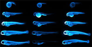
Representative images of ApoB-LP localization in zebrafish across developmental, genetic, pharmacological and dietary manipulations.
Credit: Figure 5.D of The LipoGlo reporter system for sensitive and specific monitoring of atherogenic lipoproteins by James Thierer, Stephen C. Ekker and Steven A. Farber.
Article licensed under Creative Commons Attribution 4.0 International License.

Cardiovascular diseases, or CVDs, are collectively the most notorious gang of cold-blooded killers threatening human lives today. These unforgiving villains, including the likes of coronary heart disease, cerebrovascular disease and pulmonary embolisms, are jointly responsible for more deaths per year than any other source, securing their seat as the number one cause of human mortality on a global scale.

One of the trademarks of most CVDs is the thickening and stiffening of the arteries, a condition known as atherosclerosis. Atherosclerosis is characterized by the accumulation of cholesterol, fats and other substances, which together form plaques in and on the artery walls. These plaques clog or narrow your arteries until they completely block the flow of blood, and can no longer supply sufficient blood to your tissues and organs. Or the plaques can burst, setting off a disastrous chain reaction that begins with a blood clot, and often ends with a heart attack or stroke.

Given the global prevalence and magnitude of this problem, there is a significant and urgent demand for better ways to treat CVDs. In a recent study published in Nature Communications, researchers at the Carnegie Institution for Science, Johns Hopkins University and Mayo Clinic are taking the fight to CVDs through the study of low-density lipoproteins (LDLs), the particles responsible for shuttling bad cholesterol throughout the bloodstream.

Continue reading “Striking Fear into the Heart of Cardiovascular Disease Using Zebrafish and NanoLuc® Luciferase”

Manipulating Koala Microbiomes Using…Poo Pills?

“Koalas – mum and baby” by Amanda Penrose is licensed under CC BY-NC-SA 2.0 

In recent years, it’s become a well-documented fact that koalas are about as picky as they are adorable. These beloved Australian marsupials have evolved to become ecological specialists: consumers that feed primarily on a single organism, or small number of organisms. Eucalyptus, their organism of choice, encompasses approximately 900 species, most of which are native to Australia. To the koala’s benefit, the leaves of eucalyptus trees are difficult to digest, low in protein content and their chemical composition contains compounds that are toxic. This makes their competition for eucalyptus with other species virtually nonexistent.

That’s not to say there isn’t competition amongst themselves. Of those 900 species of eucalyptus, koalas are only really known to feed on about 40–50 of them, and of those 40–50, they tend to limit their diet to around 10. Depending on their location, however, some koalas will only stick to one preferred type, which can lead to trouble.  

Continue reading “Manipulating Koala Microbiomes Using…Poo Pills?”

The Simplex Things In Life: Utilizing Artificial Intelligence Models to Better Understand Autism

Autism Spectrum Disorder, or ASD, is nothing if not unique.

The way ASD manifests itself in people is unique; although it most often presents as some form of variable impairment in social interaction and communication, each individual has behaviors and habits that are as unique to them as snowflakes are to one another.

ASD has also proven itself to be a uniquely challenging disorder to study. In the past decade, de novo (new) mutations have been identified as key contributors to causality of ASD. However, the majority of these identified de novo mutations are located in protein-coding genes, which comprise only 1–2% of the entire human genome.

Up to this point, a majority of previous research has focused on identifying mutations located in the 20,000 identified genes in the protein-coding region, which would seem like a promising approach. Genes are the genetic blueprints for creating proteins, which control and perform crucial tasks in our bodies, such as fighting off infections, communicating between your organs, tissues, and cells as chemical messengers, and regulating your blood sugar levels. It seems like basic math: Genes + Mutations = Mutated Proteins. Mutated Proteins = Disrupted Protein Function.

However, it has been observed that all the known genes that are ASD-associated can explain only a minor fraction of new autism cases, and it is estimated that known de novo mutations in the protein-coding region contribute to not more than 30% of cases for individuals who have no family history of autism (better known as simplex ASD). This provides evidence to suggest mutations contributing to autism must additionally occur elsewhere in the genome. Continue reading “The Simplex Things In Life: Utilizing Artificial Intelligence Models to Better Understand Autism”

Useful or Useless: Weird Things Packed in Our Evolutionary Suitcase

Genetics are a curious thing. Don’t get me wrong, on paper and in theory, the study and science behind our inheritance completely checks out. However, in practice, it can still be a bit disconcerting to look in the mirror one day and recognize your father’s nose and eyebrows in your own face, or to realize you gesticulate in the same animated fashion as your mother, and sometimes hear her laugh come bubbling out of your own mouth.

More curious still are the structures and behaviors that have been carried throughout evolution to the modern era of humanity, though we are considerably distinguishable from our more primitive ancestors.

And perhaps most curious of all, are the structures we continue to pack along with us, as that have little to no known useful function in the contemporary human body. These features are better known as vestigial structures, and are classically defined as features and behaviors that no longer serve the function and purpose they were designed to perform (in comparison to other creatures with the same parts).

Currently, as I recover from the aftermath of a painful encounter with one of my own vestigial organs, I find myself considering if my late appendix ever did anything much for me, or if it’s only purpose was to lie in wait as a metaphorical ticking time-bomb. Prior to my surprise appendectomy, I hadn’t spared much thought for my appendix, and decided I wanted to honor it’s memory by learning more about it, in addition to several of our other human evolutionary leftovers. Man, I wish I would’ve asked the doctors to hang on to that bad boy for me!

The Evolutionary Junk in Our Trunk

Appendix

The appendix is perhaps the most widely known vestigial organ in the human body of today. If you’ve never seen one, the appendix is a small, pouch-like tube of tissue that juts off the large intestine where the small and large intestines connect. By comparison, in herbivorous vertebrates the appendix is much larger, and functions primarily to aid in the breakdown of cellulose in consumed plants. Today, the appendix is considered a small leftover from one of our plant-eating ancestors. As our diets have changed over time, the role our appendix plays in digestion has declined, leaving plenty of room for speculation regarding what purpose it serves now. Continue reading “Useful or Useless: Weird Things Packed in Our Evolutionary Suitcase”

When Good Proteins Go Bad

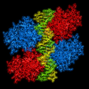
Ribbon model of p53 protein bound to DNA molecule.

Following what feels like an exceptionally long and brutal winter, I for one couldn’t be happier about the arrival of Spring and the way it makes everything seem brighter and brand-new. Soaking in the soul-warming sunshine. Reveling in the sweet melody of chirping birds. Watching the earth literally coming alive again with greenery. And for those of us who love and are enthralled by scientific discoveries like myself, the report of a recent shiny new discovery in the world of cancer research is equally as day-brightening and spirit-lifting.

To suppress tumors or to not suppress tumors: that is the question.

In the world of oncology, the protein known as p53 has long proven itself to be a primary target of interest. p53 operates as a tumor suppressor protein, often lauded as the “guardian of the human genome”, due to its dedication to governing controlled cell division and assessing damaged DNA. There are a number of cellular stressors that can wreak havoc on your DNA, including exposure to ultraviolet light or radiation, oxygen deficiency (hypoxia), and contact with hazardous chemicals.

Consider a normal-functioning p53 protein as the quality control person in a production factory. The p53 protein evaluates the products, DNA, coming down the line and determines an appropriate course of action for those that do not meet the quality standards.

Let’s say some less-than-quality DNA comes down the pipe. If the DNA is not too severely injured, p53 will alert and activate additional genes to repair the damage. However, if the products coming through are too marred to repair, p53 will shut down the whole factory, if you will, by signaling for the cell to self-destruct via apoptosis. In doing so, p53 effectively impedes tumor development by inhibiting the ability for flawed DNA to further divide.

So, it would seem like p53 has proven itself to be an undeniably upstanding citizen of the protein variety, right? The unfortunate truth of the matter is p53 balances delicately on a double-edged sword, establishing itself as the veritable Dr. Jekyll and Mr. Hyde of the cellular world: usually unquestionably good, but sometimes unspeakably evil. Continue reading “When Good Proteins Go Bad”