Scientific progress is iterative. Small advances and bits and pieces of knowledge are accumulated over time with each advance depending on the body of work that has preceded it. Being able to replicate the work from one step to another in this march of science is critical, and it is not easy. We all recognize that a well-documented procedure goes a long way toward enabling reproducibility, but the procedure is not the only hurdle to reproducibility, and it’s not even the main one.
Many of the reagents researchers use are derived from or produced by living organisms. A decade ago, in 2015, an analysis of preclinical research estimated that irreproducibility costs the U.S. around $28 billion a year. When the authors traced the causes, the largest single category wasn’t study design or data analysis. It was biological reagents and reference materials (1).
When Bill Linton first developed the idea of a company that supports scientists in the 1970s, his question was: “What can we do to make your work easier?” The answer he kept getting was the same: researchers wished they could buy reliable restriction enzymes instead of purifying their own for every experiment. It would reduce variability and enable them to focus on the interesting biological questions rather than the requisite protein isolations needed to ask those questions.
Biotechnology and life science research have come a long way since then. We now take reliable, commercially available restriction enzymes for granted. Still, Promega keeps asking Bill’s original question: What can we do to make your work easier? The answers to that question have led to ground-breaking luciferase reporter technology, add-mix-measure assays for everything from cell viability to P450 activity to NADP/H detection, and standardized thaw-and-use primary cell lines for bioassays.
Today’s Difficult Reagent: Primary Antibodies
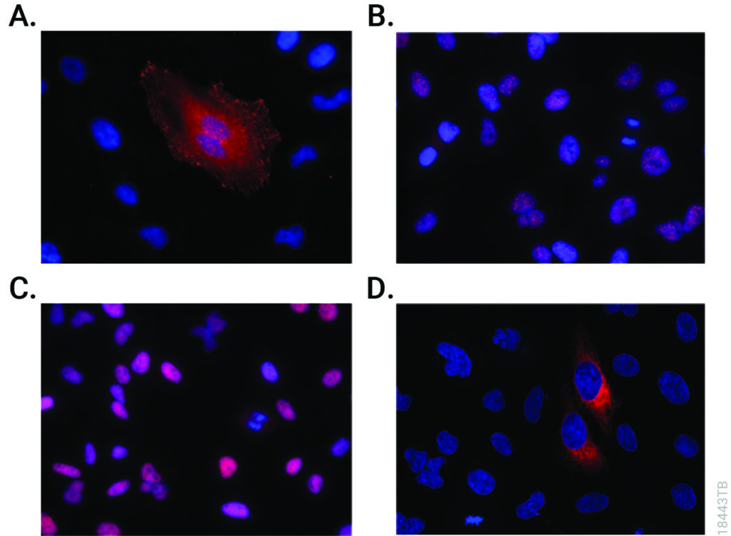
Among the most difficult reagents to work with and standardize are antibodies. If you are generating polyclonal primary antibodies that are not commercially available to study your target protein, you first need to find an animal with clean pre-immune serum. Usually that serum is evaluated by Western blot, which itself is an inherently fussy technique. And polyclonal antibodies are just that—polyclonal. What is generated after exposure to your antigen is a mixture of antibodies, and the composition of the mixture can change over time as the animal’s immune response matures. This gives you batch-to-batch variability and can make reproducibility more difficult. Commercially produced polyclonal antibodies are subject to the same exact issues. In August 2026, a news article in Nature reported that a metascientist had released a database of more than 18,000 questionable validation images across the catalogs of 15 antibody suppliers. All reflect the antibodies may not be performing like the scientists expect (Garisto 2026). Additionally, production of antibodies requires animal resources. High-quality, application validated antibodies are expensive as well. Using primary antibodies to study proteins is a significant pain point for the bench scientist.
It’s Not Just the Antibodies. It’s the Western Blots Too.
Tagged proteins or high-abundance exogenous expression of a target protein will not always reflect the endogenous protein behavior, localization or function. Large tags can interfere with folding and function; over expression of a protein can produce false protein interactions. Your results could be showing you artifacts resulting from a situation in your cells that is simply not biologically relevant.
If you are studying your target proteins (and other molecules) using Western blotting, post-translational modifications such as phosphorylation, ubiquitination, and cleavage shifts can complicate the interpretation of your results. Multiple bands on a blot can be hard to understand—is the protein cleaved? Is there cross-reactivity with your antibody to another protein? Is your sample degraded?
Continue reading “What Can We Do to Make Your Work Easier? Addressing the Challenges of Working with Primary Antibodies”