Determination of Antibody Mechanism of Action Using IdeS Protease

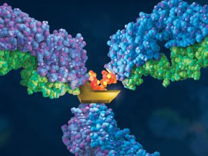
Monoclonal antibodies (mAbs) have been widely used to eliminate undesired cells via various mechanisms, including antibody-dependent cell-mediated cytotoxicity (ADCC), complement-dependent cytotoxicity (CDC) and programmed cell death (PCD). Unlike the Fc-dependent mechanism of ADCC and CDC, certain antibody–antigen interactions can evoke direct PCD via apoptosis or oncosis. Previously, researchers have reported the specific killing of undifferentiated human embryonic stem cells (hESC) by mAb84 (IgM) via oncosis (1)

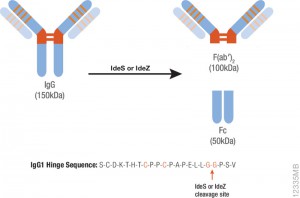
In a recent publication (2), a monoclonal antibody (mAb), TAG-A1 (A1), was generated to selectively kill residual undifferentiated human embryonic stem cells (hESC). One of the many experimental tools used to characterize the mechanism of oncosis was the fragmention of the A1 antibody with IdeS and papain.

Learn more about IdeS and IdeZ Protease available from Promega.

Papain digestion of IgG produces Fab fragments in the presence of reducing agent. F(ab)2 fragments of A1 were produced using IdeS Protease.

The results indicate that both Fab_A1 and F(ab)2_A1 bind to hESC but only F(ab)2_A1 retained hESC killing. Hence bivalency, but not Fc-domain, is essential for A1 killing on hESC.

  1. Choo, A.B. et al. (2008) Selection against undifferentiated human embryonic stem cells by a cytotoxic antibody recognizing podocalyxin-like protein-1. Stem Cells  26, 1454.
  2. Zheng, J.Y. et al. (2017) Excess reactive oxygen species production mediates monoclonal antibody-induced human embryonic stem cell death via oncosis. Cell Death and Differentiation 24, 546–58.

Are you looking for proteases to use in your research?
Explore our portfolio of proteases today.

Improved Method for the Rapid Analysis of Monoclonal Antibodies Using IdeS

ides_ab

Therapeutic monoclonal antibodies (MAbs) are inherently heterogeneous due to a wide range of both enzymatic and chemical modifications, such as oxidation, deamidation and glycosylation which may occur during expression, purification or storage. For identification and functional evaluation of these modifications, stability studies
are typically performed by employing stress conditions such as exposure to chemical oxidizers, elevated pH and temperature.

To characterize MAbs, a variety of analytical techniques are chosen, such as size exclusion chromatography and ion exchange chromatography. However, due to the large size of the intact MAbs, these methods lack structural resolution. Often, the chromatographic peaks resolved by SEC and IEC methods are collected and further analyzed by peptide mapping to obtain more detailed information. Peptide mapping, in which antibodies are cleaved into small peptides through protease digestion followed by LC–MS/MS analysis, is generally the method of choice for detection and quantitation of site-specific modifications. However sample preparation and lengthy chromatographic separation make peptide mapping impractical for the analysis of large numbers of samples. In contrast to peptide mapping analysis, the middle-down approach offers the advantage of high-throughput and specificity for antibody characterization.

Limited proteolysis of IgG molecules by the IdeS enzyme has been introduced for antibody characterization due to its high cleavage specificity and simple digestion procedure.

Continue reading “Improved Method for the Rapid Analysis of Monoclonal Antibodies Using IdeS”

IdeZ Protease: A New Tool for the Characterization of Antibodies

12335MB

Therapeutic monoclonal antibodies are large, complex molecules that undergo numerous post translational modifications (PTMs).  In-depth characterization of antibody PTMs remains a significant hurdle because their large size (~150 kDa) makes mass spectrometry analysis extremely challenging.

IdeS protease specifically cleaves IgGs into Fab and Fc fragments. This enzyme is highly specific and cleaves human IgG specifically at one site in the lower hinge region.  Because of the exquisite specificity of the enzyme, it produces highly homogeneous Fc and Fab fragments which are then readily analyzed using techniques such as mass spectrometry or HPLC.

Are you looking for proteases to use in your research?
Explore our portfolio of proteases today.

One of the drawbacks of IdeS is that it exhibits poor activity against mouse IgGs. IdeZ Protease is an immunoglobulin-degrading enzyme from Streptococcus equi subspecies zooepidemicus. It is an engineered recombinant protease overexpressed in E. coli. Like IdeS Protease, IdeZ Protease specifically cleaves IgG molecules below the hinge region to yield F(ab′)2 and Fc fragments.  Reduction of the digestion products produces three fragments of ~25kDa that are readily analyzed by LC-MS.

One of the key advantages of the IdeZ Protease is that it has significantly improved activity against mouse IgG2a and IgG3 subclasses compared to IdeS Protease. IdeZ Protease does not cleave mouse IgG1 or IgG2b.

Key technical parameters when digesting mouse IgGs utilizing IdeZ are the following:

• Add 1 unit of IdeZ Protease per 1µg of IgG to be digested.
• IdeZ Protease is most active in buffers at or near neutral pH. The recommended digestion buffer is 50mM sodium phosphate, 150mM NaCl (pH 6.6).
• Mouse IgG2a and IgG3 typically require 2–4 hours at 37°C  for complete digestion.
• IdeZ Protease has a histidine tag for easy removal if so desired.