Following what feels like an exceptionally long and brutal winter, I for one couldn’t be happier about the arrival of Spring and the way it makes everything seem brighter and brand-new. Soaking in the soul-warming sunshine. Reveling in the sweet melody of chirping birds. Watching the earth literally coming alive again with greenery. And for those of us who love and are enthralled by scientific discoveries like myself, the report of a recent shiny new discovery in the world of cancer research is equally as day-brightening and spirit-lifting.
To suppress tumors or to not suppress tumors: that is the question.
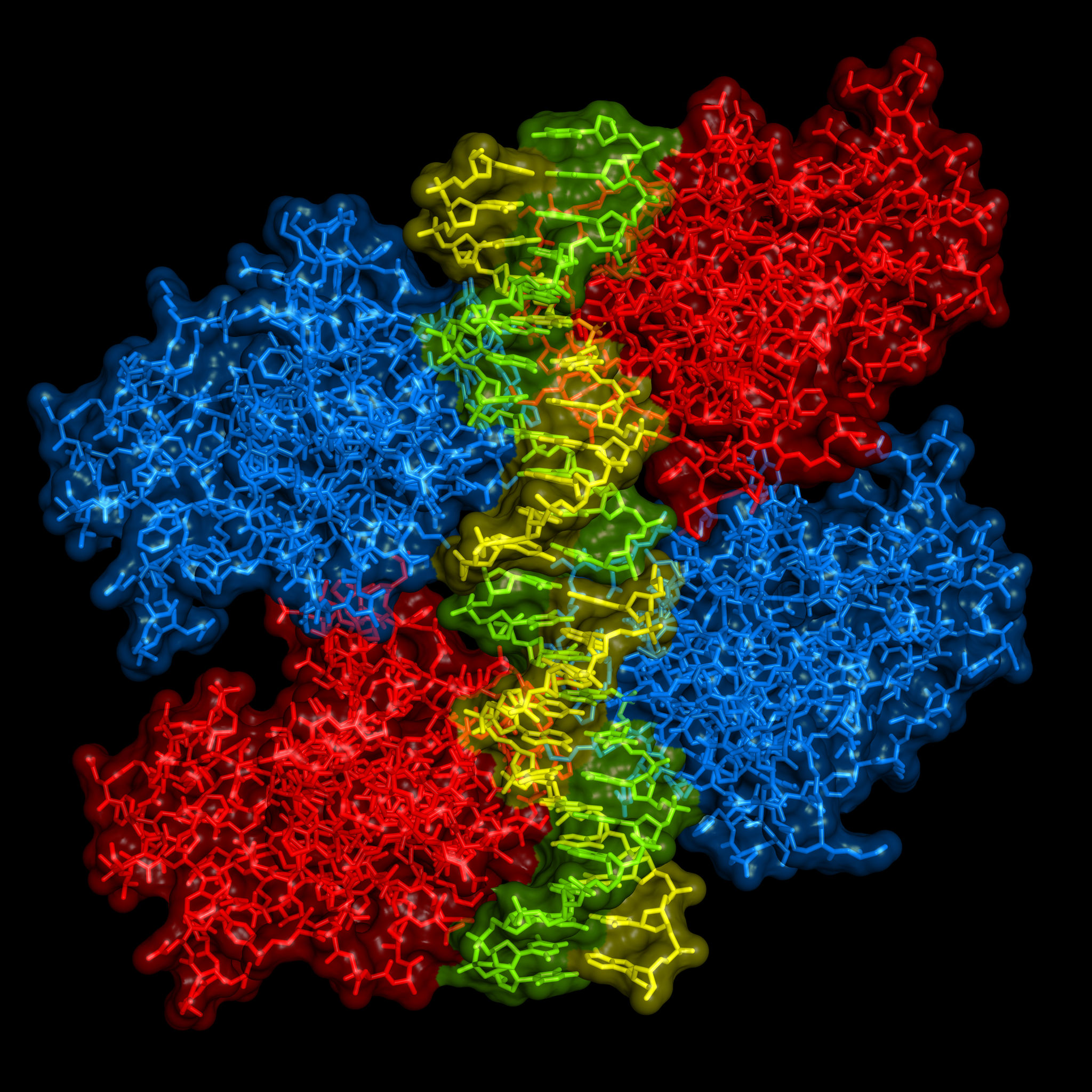
In the world of oncology, the protein known as p53 has long proven itself to be a primary target of interest. p53 operates as a tumor suppressor protein, often lauded as the “guardian of the human genome”, due to its dedication to governing controlled cell division and assessing damaged DNA. There are a number of cellular stressors that can wreak havoc on your DNA, including exposure to ultraviolet light or radiation, oxygen deficiency (hypoxia), and contact with hazardous chemicals.
Consider a normal-functioning p53 protein as the quality control person in a production factory. The p53 protein evaluates the products, DNA, coming down the line and determines an appropriate course of action for those that do not meet the quality standards.
Let’s say some less-than-quality DNA comes down the pipe. If the DNA is not too severely injured, p53 will alert and activate additional genes to repair the damage. However, if the products coming through are too marred to repair, p53 will shut down the whole factory, if you will, by signaling for the cell to self-destruct via apoptosis. In doing so, p53 effectively impedes tumor development by inhibiting the ability for flawed DNA to further divide.
So, it would seem like p53 has proven itself to be an undeniably upstanding citizen of the protein variety, right? The unfortunate truth of the matter is p53 balances delicately on a double-edged sword, establishing itself as the veritable Dr. Jekyll and Mr. Hyde of the cellular world: usually unquestionably good, but sometimes unspeakably evil.
Betrayal, thy name is p53.
As you are now fully aware, a typical p53 protein does an excellent job protecting the body from developing cancer. However, when p53 protein becomes mutated, it transforms into a protein version of the nefarious Mr. Hyde, converting from a guardian against uncontrolled cell division to a harbinger of it.
Researchers have long been aware that this betrayal by p53 plays a role in a wide variety of cancers. It has been observed that mutated p53 proteins contribute to 20 to 40 percent of breast cancers and almost 50 percent of lung cancer, ovarian cancer, and squamous cell carcinomas of the head and neck.
With statistics like that, it’s no wonder that p53 has been a target of extensive cancer studies for several decades already. In spite of these years of dedicated research, there are still no developed drugs that specifically target this obvious cancer contributor.
This is due in large part to the fundamental question that has continued to elude scientists: what causes p53 to transform from good to bad?
This above all: to thine dedicated, peer-reviewed research be true.
A team of scientists at the University of Wisconsin-Madison recently published a paper in Nature Cell Biology, that finally offers some insight into just how p53 abandons it’s good-guy persona and forsakes the very cells it is designed to protect.
The research team, comprised of Richard Anderson, Dr. Vincent Cryns, and postdoctoral fellows Mo Chen and Suyong Choi, discovered an unprecedented molecular mechanism by which mutated p53 promotes cancer.
In their research, they found the surprising involvement of an enzyme called PIPK1-α and a lipid messenger dubbed PIP2, in the regulation of mutated p53 proteins. PIPK1-α and PIP2 are typically found primarily in the cellular membrane.
In this molecular mechanism, they found that PIPK1-alpha enzyme relocates to the nucleus and joins with p53 to generate PIP2 lipid messengers when the cell undergoes stress. PIP2 also strongly associates with p53, and affixes p53 with a tag which marks p53 for additional interaction with small heat shock proteins, which stabilize the mutant p53. Once stabilized, mutant p53 is able to accumulate in the nucleus, inhibiting normal p53 function and setting the stage for rampant cell proliferation which promotes the formation of tumors.
They were also able to further demonstrate that when the PIP2 pathway was disrupted, the mutant p53 did not accumulate and cause damage. This notably implicates the pathway as an auspicious target to exploit in future development of cancer therapeutics.
The discovery of this pathway doesn’t merely unlock the door for potential drug development, it virtually kicks it open. Now that the enablers of mutant p53 have been identified, that establishes actual, tangible molecular targets that further research can hone in on.
It’s the biological equivalent of police identifying criminal suspects—it’s a lot easier to get to the bottom of a crime if you know who you’re looking for.