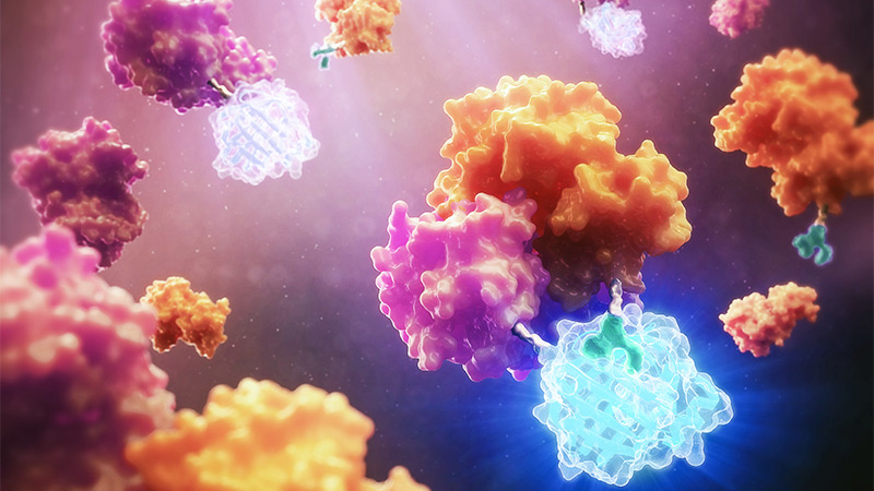
The development of NanoLuc® luciferase technology has provided researchers with new and better tools to study endogenous biology: how proteins behave in their native environments within cells and tissues. This small (~19kDa) luciferase enzyme, derived from the deep-sea shrimp Oplophorus gracilirostris, offers several advantages over firefly or Renilla luciferase. For an overview of NanoLuc® luciferase applications, see: NanoLuc® Luciferase Powers More than Reporter Assays.
The small size of NanoLuc® luciferase, as well the lack of a requirement for ATP to generate a bioluminescent signal, make it particularly attractive as a reporter for in vivo bioluminescent imaging, both in cells and live animals. Expression of a small reporter molecule as a fusion protein is less likely to interfere with the biological function of the target protein. NanoLuc® Binary Technology (NanoBiT®) takes this approach a step further by creating a complementation reporter system where one subunit is just 11 amino acids in length. This video explains how the high-affinity version of NanoBiT® complementation (HiBiT) works:
Tiny Tags to Track Viruses
An early application of NanoLuc®-based in vivo imaging was the study of viral infections. In 2013, Tran et al. reported the development of a replication-competent reporter virus for mouse model studies of influenza A (1). Previous bioluminescence imaging techniques often resulted in reporter viruses that were unstable and suffered from limited sensitivity in vivo. The NanoLuc® reporter virus exhibited pathogenicity and lethality in mice that was identical to the native influenza A virus. Using the reporter virus, the researchers were able to track viral load and dissemination in the lungs. Their results demonstrated the utility of NanoLuc® reporter viruses in studying emerging viruses and the potential impact of antiviral therapy.
A recent study demonstrated an approach to constructing reporter viruses that further improved on stability and offered greater flexibility in reporter design (2). In this study, researchers inserted the HiBiT tag into a modified oncolytic adenovirus genome, under the control of the viral E3 promoter. This promoter is activated by the viral E1A protein soon after infection. To demonstrate the stability and functionality of the reporter virus, the researchers infected both adherent and suspension prostate cancer cell lines expressing the complementary LgBiT peptide. They observed successful reconstitution of NanoBiT® luciferase activity 24 hours after infection in all cell lines tested, using the Nano-Glo® substrate.
For in vivo imaging, they monitored LgBiT-expressing cells for stable infection with the reporter virus and then infected tumors implanted in mice with either the reporter virus or a control. Intraperitoneal (IP) administration of substrate showed successful infection and good substrate distribution, leading to sustained bioluminescent emission. The researchers used a derivative of furimazine in these experiments (hydrofurimazine) due to its increased solubility and the consequent ease of IP administration at the concentrations required for imaging. This substrate, along with fluorofurimazine—which provides an even brighter in vivo signal—was developed recently for in vivo imaging applications in mice (3). These novel substrates open up new options for advancing in vivo studies with NanoLuc® reporter technologies.
Further studies of infection dynamics (2) showed that the oncolytic virus did not reach the entire tumor mass; however, this result was consistent with oncolytic adenovirus studies in clinical trials. The authors concluded that their approach is applicable to studying a wide range of viruses, especially those unable to tolerate large transgene insertions.
Better with BRET: Antibody-Receptor Interactions
One challenge with using bioluminescence for imaging of solid tumors or other tissues is the emission wavelengths of the bioluminescent reaction. Hemoglobin absorbs primarily in the blue and green regions of the visible spectrum; therefore, an ideal bioluminescent reaction would emit light in the far red region (4). NanoLuc® luciferase reactions emit light at 460nm, in the blue region. Fluorescent dyes with suitable emission wavelengths traditionally have been used for in vivo imaging but are limited by the requirement for an external excitation source.
Bioluminescence resonance energy transfer (BRET) offers the best of both worlds: the small size of a NanoLuc® luciferase tag and the brightness of a fluorescent molecule. In BRET, energy is transferred from a donor luciferase substrate to a fluorescent acceptor molecule, when the two are brought into close proximity. The fluorescent molecule then re-emits light at its own optimal wavelength. Because NanoLuc® luciferase is ATP-independent, it can also be used in BRET applications where the reporter is expressed outside the cell, creating new possibilities for studying molecular interactions that occur on the cell surface.
Tang et al. used BRET to study the binding of therapeutic antibodies to receptors in live animals (5). They fused NanoLuc® luciferase to the N-terminus of the epidermal growth factor receptor (EGFR), and used the fluorescent dye DY605 covalently attached to the therapeutic antibody cetuximab as the acceptor. While addition of furimazine to the NanoLuc®-EGFR fusion protein alone resulted in the expected emission peak at 460nm, antibody binding produced an additional peak at 625nm. The separation between these two emission peaks was sufficient for reliable and sensitive detection, as characterized in cell culture.
For in vivo experiments, the researchers examined the receptor occupancy and pharmacokinetics of cetuximab using a tumor xenograft model in nude mice. They observed incomplete receptor occupancy, even at higher than therapeutic doses of cetuximab, indicating that only a fraction of receptors were available for antibody binding. Despite some technical limitations, BRET proved to be a useful, noninvasive imaging technique to establish dose-response relationships for cetuximab binding to EGFR. The authors note that the method can be generalized to studying the efficacy and toxicity of other therapeutic antibodies.
An alternative approach to BRET is to fuse a bioluminescent protein with a fluorescent one. An example is the development of the Antares reporter system, in which NanoLuc® luciferase is fused to two copies of cyan-excitable orange fluorescent protein 1 (Cy-OFP1). This approach is summarized in Making BRET the Bright Choice for In vivo Imaging: Use of NanoLuc® Luciferase with Fluorescent Protein Acceptors. Su et al. (3) used the Antares reporter together with a firefly luciferase reporter for in vivo imaging of two cell populations in mice. They were able to measure tumor size and also visualize chimeric antigen receptor (CAR) T-cell migration in the same animal.
For more in vivo imaging applications of NanoLuc® luciferase, or to inquire about new detection solutions, see our NanoLuc® Bioluminescence Imaging resource page.
References
- Tran, V. et al. (2013) Highly sensitive real-time in vivo imaging of an influenza reporter virus reveals dynamics of replication and spread. J. Virol. 87, 13321.
- Gaspar, N. et al. (2020) NanoBiT system and hydrofurimazine for optimized detection of viral infection in mice—a novel in vivo imaging platform. Int. J. Mol. Sci. 21, 5863.
- Su, Y. et al. (2020) Novel NanoLuc substrates enable bright two-population bioluminescence imaging in animals. Nat. Methods 17, 852–860.
- Zhao, H. et al. (2005) Emission spectra of bioluminescent reporters and interaction with mammalian tissue determine the sensitivity of detection in vivo. J. Biomed. Optics 10(4), 041210.
- Tang, Y. et al. (2019) A bioluminescence resonance energy transfer-based approach for determining antibody-receptor occupancy in vivo. iScience 15, 439–451.
Related Posts
Latest posts by Ken Doyle (see all)
- Will Artificial Intelligence (AI) Transform the Future of Life Science Research? - February 1, 2024
- RAF Inhibitors: Quantifying Drug-Target Occupancy at Active RAS-RAF Complexes in Live Cells - September 5, 2023
- Synthetic Biology: Minimal Cell, Maximal Opportunity - July 25, 2023
