When he was a kid, Matt Hanson would disappear into the basement for an entire day and emerge later with a completed model of the USS Constitution or a completed robot or a new rocket (he still makes model rockets). Design and how things fit together have always fascinated him, so a career in science was a natural fit as well.
Today Matt is a Quality Control Supervisor/QA Senior Scientist at Promega Corporation at the Madison, WI, USA, campus. He has been with Promega for 5 years now.
After completing his undergraduate studies in molecular biology, a masters in zoology where he focused on cell biology, and a PhD in developmental biology and immunology, Matt was fortunate to pursue a successful and rewarding career as an Associate Staff Scientist in the Department of Surgery at the University of Wisconsin-Madison. His work focused on diabetes and transplantation biology.
So why did Matt join the scientific staff at Promega?
Electrophysiology experiments provide a view into the cell with amazing detail. The paper reviewed here describes a molecular reporter biosensor (NanoBRET) that can offer the same kind of temporal and spatial resolution traditionally reserved for extremely labor-intensive experiments like patch clamp analysis.
I confess that I struggled through biophysics, and my Bertil Hille textbook Ion Channels of Excitable Membranes lies neglected somewhere in a box in my basement (I have not tossed it into the recycle bin—I can’t bear too, I spent too much time bonding with that book in graduate school).
My struggles in that graduate class and my attendance at the seminars of my grad school colleagues who were conducting electrophysiological studies left me with a sincere awe and appreciation of both the genius and the artistry required to produce nice electrophysiology data. The people who are good at these experiments are artists—they have the golden touch when it comes to generating that megaohm seal between a piece of cell membrane and a finely pulled glass pipette. And, they are brilliant scientists, they really understand the physics, the chemistry and the biology of the cells they study from a perspective that very few scientists ever develop.
Electrophysiology data, which often demonstrate the gating of a single channel protein in response to a single stimulus in real time–ions crossing a membrane through a single protein–are amazing for their ability, unlike virtually any other experimental data for the story they can tell about what is going on in a cell in real time under physiological conditions.
When constructs were ectopically expressed in HEK 293T/17 cells, we obtained very similar kinetics for the GPCR-driven responses between NanoBRET™ biosensors and the patch clamp recordings.
They continue:
Indeed, the activation rates that we observed were very similar to those of GPCR-stimulated GIRKs [G protein-coupled, inwardly rectifying K+ channel] in native cells, suggesting that the conditions of this assay closely match the in vivo setting. This finding further demonstrates the ability of the system to resolve the fast, physiological relevant kinetics of GPCR signaling.
Over the last few months we have published several blogs about qPCR—from basic pointers on avoiding contamination in these sensitive reactions to a collection of tips for successful qPCR. Today we look in depth at a paper that describes the design and and optimization of a qPCR assay, and in keeping with the season of winter in the Northern hemisphere, it is only fitting that the assay tests for the abundance and identity of ice-nucleating bacteria.
Ice-nucleating bacteria are gram-negative bacteria that occur in the environment and are able to “catalyze” the formation ice crystals at warmer temperatures because of the expression of specific, ice-nucleating proteins on their outer membrane. Ice-nucleating bacteria are found in abundance on crop plants, especially grains, and are estimated to cause one-billion dollars in crop damage from frost in the United States alone.
In addition to their abundance on crop plants, ice-nucleating bacteria are also found on natural vegetation and have been isolated from soil, snow, hail, cloud water, in the air above crops under dry conditions and during rain fall. They have even been isolated from soil, seedlings and snow in remote locations in Antarctica. For the bacteria, ice nucleation may be a method to promote dissemination through rain and snow.
Although ice-nucleating bacteria have been isolated from clouds, ice and rain, little is known about their true contribution to precipitation or other events such as glaciation. Are such bacteria the only source of warm-temperature (above temperatures at which ice crystals form without a catalyst) ice nucleation? Can they trigger precipitation directly? What are the factors that trigger their release from vegetation into the atmosphere? Can we determine their abundance and variety in the environment?
For three out of the last four years, we have been honored to have one of our key technologies named a Top 10 Innovation by The Scientist. This year the innovative NanoBiT™ Assay (NanoLuc® Binary Technology) received the recognition. NanoBiT™ is a structural complementation reporter based on NanoLuc® Luciferase, a small, bright luciferase derived from the deep sea shrimp Oplophorus gracilirostris.
Using plasmids that encode the NanoBiT complementation reporter, you can make fusion proteins to “report” on protein interactions that you are studying. One of the target proteins is fused to the 18kDa subunit; the other to the 11 amino acid subunit. The NanoBiT™ subunits are stable, exhibiting low self-affinity, but produce an ultra-bright signal upon association. So, if your target proteins interact, the two subunits are brought close enough to each other to associate and produce a luminescent signal. The strong signal and low background associated with a luminescent system, and the small size of the complementation reporter, all help the NanoBiT™ assay overcome the limitations associated with traditional methods for studying protein interactions.
The small size reduces the chances of steric interference with protein interactions. The ultra bright signal, means that even interactions among proteins present in very low amounts can be detected and quantified–without over-expressing large quantities of non-native fusion proteins and potentially disrupting the normal cellular environment. And the NanoBiT™ assay can be performed in real time, in live cells.
The NanoBiT™ assay is already being deployed in laboratories to help advance understanding of fundamental cell biology. You can see how one researcher is already taking full advantage of this innovative technology in the video embedded below:
Visit the Promega web site to see more examples more examples how the NanoBiT™ assay can break through the traditional limitations for studying protein interactions in cells.
You can read the Top 10 article in The Scientisthere.
Often a diagnosis of thyroid cancer is associated with a good prognosis and fairly straightforward surgical treatments to remove the tumor followed by radioactive iodine ablation. Such treatment works well in tumors that have not metastasized and retain enough of their thyroid cell “identity” that they can still accumulate radioactive iodine.
However, aggressive thyroid cancers, which often metastasize and recur, do not respond to standard treatments because they are generally too dedifferentiated to accumulate iodine, so alternative treatments are needed.
One approach is to look for compounds that will reverse dedifferentiation, making tumor cells more likely to take up and concentrate radioactive iodine regardless of their location in the body. One possible target to effect dedifferentiation is epigenetic modification of histone proteins.
Histone proteins are more than the structural components of the nucleosome that organizes the chromatin inside cells. Histone proteins are subject to a host of protein modifications on their N-terminal tails such as acetylation, phosphorylation, methylation, ubiquitination and ADP-ribosylation. These various modifications are seen as creating a “histone code” that is read by other proteins and protein complexes (1). This code regulates patterns of gene expression and activity for a cell—in part resulting in a differentiated phenotype. Previous studies have suggested that some histone deacetylase (HDAC) inhibitors (e.g., valproic acid) can reverse some of the dedifferentiation associated with aggressive cancers (2).
This week’s video takes us to a forest fire on Mauna Kea, a dormant volcano on the island of Hawaii. One of the firefighters captured this amazing video of a fire whirl that erupted as the air temperature near the ground grew very hot. Fire whirls like this one are caused by extreme heat rising from the ground rather than a confluence of atmospheric events, but they can be be every bit as destructive as atmospheric tornadoes and cause a forest fire to continue to burn out of control.
There are written records of fire tornadoes including several that developed after lightning struck an oil storage facility near San Luis Obispo, CA, USA in 1926. In 2003, scientists confirmed true fire tornadoes in Australia associated with the Canberra fires. In this case the fire tornadoes produced damage consistent with the intensity of an F2 tornado. In The Great Pestigo Fire in 1871, the town of Peshtigo, WI, may well have been consumed by fire tornadoes. Dry weather conditions and slash and burn farming practices contributed to this devastating fire (as they did the more famous Chicago fire that occurred on the same day). Strong winds carried a forest fire into the mill town of Peshtigo, WI, and researchers theorize that cut timber and wooden structures of the town fueled such intense heat that a massive fire whirls formed, consuming the town. You can read some compelling stories about the Peshtigo fire here and here.
Understanding the conditions under which firestorms and fire tornadoes form hopefully will lead to a better understanding of how forest and brush fires spread and allow scientists and fire control experts to develop more effective methods of control.
Robert Hooke first coined the term “cell” after observing plant cell walls through a light microscope—little empty chambers, fixed in time and space. However, cells are anything but fixed.
Cells are dynamic: continually responding to a shifting context of time, environment, and signals from within and without. Interactions between the macromolecules within cells, including proteins, are ever changing—with complexes forming, breaking up, and reforming in new ways. These interactions provide a temporal and special framework for the work of the cell, controlling gene expression, protein production, growth, cell division and cell death.
Visualizing and measuring protein:protein interactions at the level of the cell without perturbing them is the goal of every cell biologist.
A recent article by Thomas Machleidt et al. published in ACS Chemical Biology, describes a new technology that brings us closer to being able to realize that goal.
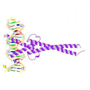
Crystal Structure of MYC MAX Heterodimer bound to DNA ImageSource=RCSB PDB; StructureID=1nkp; DOI=http://dx.doi.org/10.2210/pdb1nkp/pdb;
In 1982, picked up because of its homology to chicken virus genes that could transform cells, MYC became one of the first human genes identified that could drive cellular transformation (1,2). Since that time countless laboratories have prodded and poked the human MYC gene, the MYC protein, their homologs in other animal models, and their transforming viral counterparts.
MYC is a transcription factor and forms heterodimers with a required protein partner, MAX, before binding to the E box sequences of DNA regulatory regions (3). MYC regulates gene expression of many targets through interactions with a host of proteins, often referred to as the MYC Interactome (2). In fact, MYC is estimated to bind 10–15% of the genome, and it regulates the expression of genes that are transcribed by by each of the three RNA polymerases (2).
MYC plays a central role in regulating cell growth, proliferation, apoptosis, differentiation and transformation, acting as a central integrator of cellular signals. MYC is tightly regulated at multiple levels from gene expression to protein stability. Dysregulation (usually upregulation) of the amount and stability of Myc protein is observed in many human cancers. Even in cancers in which MYC is not directly involved in transforming cells, its normal expression is often required to support the extracellular matrix and/or vascularization necessary for tumor growth and formation (4).
Because MYC is such a central player cancer pathology, it is an attractive target for cancer therapeutics (2) .
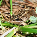
One of the hallmarks of the arrival of Spring in Wisconsin is the cacophony of evening croaks and calls from the Spring Peepers and Chorus frogs. Indeed frogs and toads are ubiquitous around the globe, and many of us who have become life scientists (even those of us who have relegated ourselves to the world of macromolecules, cell signaling networks, and nucleic acids) probably spent some time in our childhood chasing and catching frogs.
But what happens to those frogs and toads over the harsh winter months in places like Wisconsin? Well, their strategies are species-dependent, but at least some of them overwinter by freezing, and the story of one species, the Wood Frog, is quite amazing. Think about it. It freezes from the inside out. No heart beat, no circulation, completely dormant. Then in response to some unknown signal (day length? temperature? angle of the sun?), bodily functions slowly resume. What kind of cell signaling cascade controls that response?
Here is a video from NOVA about the Wood Frog and its amazing deicing event. The next time you are out on a Spring or Summer evening and you hear a chorus of frogs calling, you can think about the incredible molecular story behind the event and be even more impressed!
What will you be doing on 3/14/15 at precisely 9:26:53? Twice the clocks will align with the first few digits of my favorite irrational and transcendental number (3.141592653…) or π. Over 1 trillion digits to the right of the decimal point have been calculated, and they still go on, never making a pattern, never repeating.
Take any circle, that blueberry pie that your grandmother baked would be a good choice. Measure the circumference and divide by the diameter and you will have Pi, the mathematical constant that represents the ratio of circumference to diameter in a circle. You probably encountered Pi in your early mathematics education when learning the formula for the area of a circle (2πr2).
XWe use cookies and similar technologies to make our website work, run analytics, improve our website, and show you personalized content and advertising. Some of these cookies are essential for our website to work. For others, we won’t set them unless you accept them. To learn more about our approach to Privacy we invite you to Read More
By clicking “Accept All”, you consent to the use of ALL the cookies. However you may visit Cookie Settings to provide a controlled consent.
We use cookies and similar technologies to make our website work, run analytics, improve our website, and show you personalized content and advertising. Some of these cookies are essential for our website to work. For others, we won’t set them unless you accept them. To find out more about cookies and how to manage cookies, read our Cookie Policy.
If you are located in the EEA, the United Kingdom, or Switzerland, you can change your settings at any time by clicking Manage Cookie Consent in the footer of our website.
Necessary cookies are absolutely essential for the website to function properly. These cookies ensure basic functionalities and security features of the website, anonymously.
Cookie
Duration
Description
cookielawinfo-checbox-analytics
11 months
This cookie is set by GDPR Cookie Consent plugin. The cookie is used to store the user consent for the cookies in the category "Analytics".
cookielawinfo-checbox-functional
11 months
The cookie is set by GDPR cookie consent to record the user consent for the cookies in the category "Functional".
cookielawinfo-checbox-others
11 months
This cookie is set by GDPR Cookie Consent plugin. The cookie is used to store the user consent for the cookies in the category "Other.
cookielawinfo-checkbox-advertisement
1 year
The cookie is set by GDPR cookie consent to record the user consent for the cookies in the category "Advertisement".
cookielawinfo-checkbox-necessary
11 months
This cookie is set by GDPR Cookie Consent plugin. The cookies is used to store the user consent for the cookies in the category "Necessary".
cookielawinfo-checkbox-performance
11 months
This cookie is set by GDPR Cookie Consent plugin. The cookie is used to store the user consent for the cookies in the category "Performance".
gdpr_status
6 months 2 days
This cookie is set by the provider Media.net. This cookie is used to check the status whether the user has accepted the cookie consent box. It also helps in not showing the cookie consent box upon re-entry to the website.
lang
This cookie is used to store the language preferences of a user to serve up content in that stored language the next time user visit the website.
viewed_cookie_policy
11 months
The cookie is set by the GDPR Cookie Consent plugin and is used to store whether or not user has consented to the use of cookies. It does not store any personal data.
Analytical cookies are used to understand how visitors interact with the website. These cookies help provide information on metrics the number of visitors, bounce rate, traffic source, etc.
Cookie
Duration
Description
SC_ANALYTICS_GLOBAL_COOKIE
10 years
This cookie is associated with Sitecore content and personalization. This cookie is used to identify the repeat visit from a single user. Sitecore will send a persistent session cookie to the web client.
vuid
2 years
This domain of this cookie is owned by Vimeo. This cookie is used by vimeo to collect tracking information. It sets a unique ID to embed videos to the website.
WMF-Last-Access
1 month 18 hours 24 minutes
This cookie is used to calculate unique devices accessing the website.
_ga
2 years
This cookie is installed by Google Analytics. The cookie is used to calculate visitor, session, campaign data and keep track of site usage for the site's analytics report. The cookies store information anonymously and assign a randomly generated number to identify unique visitors.
_gid
1 day
This cookie is installed by Google Analytics. The cookie is used to store information of how visitors use a website and helps in creating an analytics report of how the website is doing. The data collected including the number visitors, the source where they have come from, and the pages visted in an anonymous form.
Advertisement cookies are used to provide visitors with relevant ads and marketing campaigns. These cookies track visitors across websites and collect information to provide customized ads.
Cookie
Duration
Description
IDE
1 year 24 days
Used by Google DoubleClick and stores information about how the user uses the website and any other advertisement before visiting the website. This is used to present users with ads that are relevant to them according to the user profile.
test_cookie
15 minutes
This cookie is set by doubleclick.net. The purpose of the cookie is to determine if the user's browser supports cookies.
VISITOR_INFO1_LIVE
5 months 27 days
This cookie is set by Youtube. Used to track the information of the embedded YouTube videos on a website.
Performance cookies are used to understand and analyze the key performance indexes of the website which helps in delivering a better user experience for the visitors.
Cookie
Duration
Description
YSC
session
This cookies is set by Youtube and is used to track the views of embedded videos.
_gat_UA-62336821-1
1 minute
This is a pattern type cookie set by Google Analytics, where the pattern element on the name contains the unique identity number of the account or website it relates to. It appears to be a variation of the _gat cookie which is used to limit the amount of data recorded by Google on high traffic volume websites.


 Often a diagnosis of thyroid cancer is associated with a good prognosis and fairly straightforward surgical treatments to remove the tumor followed by radioactive iodine ablation. Such treatment works well in tumors that have not metastasized and retain enough of their thyroid cell “identity” that they can still accumulate radioactive iodine.
 This week’s video takes us to a forest fire on Mauna Kea, a dormant volcano on the island of Hawaii. One of the firefighters captured this amazing video of a fire whirl that erupted as the air temperature near the ground grew very hot. Fire whirls like this one are caused by extreme heat rising from the ground rather than a confluence of atmospheric events, but they can be be every bit as destructive as atmospheric tornadoes and cause a forest fire to continue to burn out of control.
 One of the hallmarks of the arrival of Spring in Wisconsin is the cacophony of evening croaks and calls from the Spring Peepers and Chorus frogs. Indeed frogs and toads are ubiquitous around the globe, and many of us who have become life scientists (even those of us who have relegated ourselves to the world of macromolecules, cell signaling networks, and nucleic acids) probably spent some time in our childhood chasing and catching frogs.