The ability to isolate and assay circulating cell-free DNA from plasma holds promise for improved diagnostics and treatment in the clinic. The use of blood-based non-invasive prenatal testing (NIPT) has been well described. Such testing is based on circulating cell-free fetal DNA in blood of a pregnant woman for diagnosis and screening of chromosomal anueploidy (e.g. Trisomy 21, Down Syndrome), sex-linked diseases, and genetic diseases that are known to result from a specific mutation in a single gene (1). Additionally, most cancers carry somatic mutations that are unique to the tumors, and dying tumor cells release small pieces of their DNA into the blood stream (2). This circulating cell-free tumor DNA can be used as a biomarker to “follow” cancer progression or regression during treatment, and diagnostic methods also are being developed to detect even early stage cancers from circulating tumor DNA (3). Further, increases in circulating cell-free DNA have been well documented after intense exercise, trauma, sepsis and even associated with autoimmune diseases such as system lupus erythematosus (SLE; 1,4). In these latter examples increases in extracellular DNA are associated with evolutionarily conserved innate immune responses involving the production of neutrophil extracellular traps (NETs). Monitoring the circulating cell-free DNA of NETs has implications for treatment and diagnosis of autoimmune diseases, cardiovascular events and traumatic injuries (4–7).
How Neutrophils Weave a Defensive Web
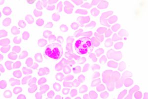
Neutrophils are the most abundant type of white blood cell and are part of the innate immune response, participating in non-specific immune responses to injury or pathogens. They are one of three types of granuolcytes, and can be recognized by their multi-lobed nucleus and the prominent granules that fill their cytoplasm. Generally they are first to the scene of injury or infection. Early in my scientific career, I was taught that neutrophils fought disease via phagocytosis and occasionally by firing a barrage of toxic enzymes and molecules at invaders. Mostly though they released cytokines that recruited the “important” cells of the specific immune system to the area.
For these reasons, I never really thought much about neutrophils. That is until recently, when I learned about Neutrophil Extracellular Traps (NETs). It turns out that neutrophils are pretty awesome, sacrificing themselves in a cloud-like explosion of DNA, chromatin, and granule proteins
Continue reading “Weaving Tangled Webs with Cell-Free DNA”
 Let’s face it, most lab techs and purchasing agents aren’t all that happy when you send them an Instagram picture of your latest lunchroom-napkin cloning strategy as your order form for your next big cloning experiment. So we have created the
  Already have a favorite vector and a freezer full of restriction enzymes? No problem, skip those steps and move on to getting the perfectly sized nucleic acid markers or the particular polymerase your experiment requires.


![Fig 4. Four point MMOA screen for tideglusib and GW8510. Time dependent inhibition was evaluated by preincubation of TbGSK3β with 60 nM tideglusib and 6 nM GW-8510 with 10μM and 100μM ATP. (A). Tideglusib [60 nM] in 10μM ATP. (B). GW8510 [60 nM] in 10μM ATP. (C.) Tideglusib [60 nM] at 100μM ATP. (D.) GW8510 [60 nM] at 100μM ATP. All reactions preincubated or not preincubated with TbGSK3β for 30 min at room temperature. Experiments run with 10μM GSM peptide, 10μM ATP, and buffer. Minute preincubation (30 min) was preincubated with inhibitor, TbGSK3β, GSM peptide, and buffer. ATP was mixed to initiate reaction. No preincubation contained inhibitor, GSM peptide, ATP, and buffer. The reaction was initiated with TbGSK3β. Reactions were run at room temperature for 5 min and stopped at 80°C. ADP formed was measured by ADP-Glo kit. Values are mean +/- standard error. N = 3 for each experiment and experiments were run in duplicates. Control reactions contained DMSO and background was determined using a zero time incubation and subtracted from all reactions. Black = 30 min preincubation Grey = No preincubation.](https://www.promegaconnections.com/wp-content/uploads/2016/04/journal.pntd_.0004506.g004-243x300.png)