Pain management, particularly long-term pain management, is a difficult problem. Opioids are effective at reducing pain, but they are addictive and carry with them the significant costs of addiction and overdose that affect not only the individual but society as well.
Recently, work presented in Science by Reeder and colleagues proposes an engineering solution to pain management: miniature, implantable devices that can deliver localized and reversible neural blocking to reduce pain. While such devices could deliver any kind of stimulus ranging from electrical to pharmacological, the researchers developed a device that temporarily blocks neural signals by cooling.
The first monoclonal antibody (mAb) was produced in a lab 1975, and the first therapeutic mAb was introduced in the United States to prevent kidney transplant rejection in 1986. The first mAb used in cancer treatment the anti-CD20 mAb, rituximab, was used to treat non-Hodgkin’s lymphoma and chronic lymphocytic leukemia. Today therapeutic mAbs have become a mainstay of cancer, autoimmune disease, and metabolic disease therapies and include HERCEPTIN® used to treat certain forms of breast cancer, Prolia used to treat bone loss in post-menopausal women, and Stelara used to treat autoimmune diseases like psoriatic arthritis and severe Crohn disease, among many others. Therapeutic mAbs bind targets with high specificity and affinity and they can recruit effector cells to drive target elimination through mechanisms such as antibody-dependent cellular cytotoxicity (ADCC) or antibody-dependent cellular phagocytosis (ADCP), making them highly specific, effective therapies.
Methylglyoxal is responsible for post translational protein modifications, that result in advanced glycation endproducts (AGEs), which are associated with aging and disease.
Post-translational modifications (PTM) of proteins are essential for the function of many proteins, but aberrant modification of protein residues also can interfere with protein function. PTMs occur in two ways. Proteins may be modified through the activity of enzymes such as kinases, phosphorylases, glycosylases and others that add or remove specific chemical moieties to amino acid residues. PTMs can also result from non-enzymatic reaction between electrophilic compounds and nucleophilic arginine and lysine residues within a protein. Metabolites and metabolic by products produced during glycolysis, especially glyoxal and methylglyoxal (MGO), are examples of such electrophilic compounds. These compounds can react with arginine and lysine to produce advanced glycation end products (AGEs), which are biomarkers associated with aging and degenerative diseases such as Alzheimer disease, diabetes and others. MGO is also elevated in tumors that have switched from oxidative phosphorylation to glycolysis as their main energy production pathway.
Only limited information is available about site-specific MGO PTMs in mammal cells, and most studies have focused on measuring the amount of MGO modifications in a treatment scenario compared to a control. Donnellan and colleagues recently published work to identify specific MGO protein modifications. They used a “bottom-up” proteomic analysis of WIL2-NS B lymphoblastoid whole-cell lysates to identify specific MGO-modified proteins. In particular, the group was looking for modifications in proteins that might explain how MGO activity contributes to aneuploidy.
For the study, 100µg of cellular protein extract was reduced with dithiothreitol and then alkylated with chloroacetamide. The sample was diluted to reduce urea concentration. Trypsin Gold was added and samples were digested for 8 hours at 37°C. Digestion was terminated by adding formic acid. For ProAlanase digestion, 20µg of protein was reduced, alkylated and diluted to reduce urea concentration before adding digesting with ProAlanase for 4 hours at 37°C.
Are you looking for proteases to use in your research? Explore our portfolio of proteases today.
The authors identified 519 MGO-modified proteins. Most of the modifications were identified in the trypsin digestion reactions; however, ProAlanase digestion increased the number of MGO modifications identified by approximately 25% (with less than 4% of the modification sites being detected in both the ProAlanase and trypsin digestion reactions. The authors suggest that ProAlanase increased sequence coverage to reveal sites not detected in the trypsin digestions. Therefore, they conclude that ProAlanase can be used along with trypsin digestion to increase the identification of MGO modifications.
ProAlanase can be used along with trypsin digestion to increase the identification of MGO modifications.
MGO-modified proteins from the WIL2-NS whole cell lysates included proteins involved in glycolysis, translation initiation, protein folding, mRNA splicing, cell-to-cell adhesion, heat response, nucleosome assembly, protein SUMOylation and the G2/M cell cycle transition. More work to further characterize the sites of these modifications and their potential effects on the function of the modified proteins is ongoing.
Donnelian, L et al. (2022) Proteomic Analysis of Methylglyoxal Modifications Reveals Susceptibility of Glycolytic Enzymes to Dicarbonyl Stress Int. J. Mol. Sci.23(7), 3689 doi.org/10.3390/ijms23073689
Vaccine research and development is a major area of focus for life scientists across the globe. Clinical trials have shown that vaccines that target tumors show promise for cancer treatment. Additionally, the emergence of new zoonotic diseases has revealed a need to develop vaccines quickly as the world becomes more global and human populations interact more often with each other and wild habitats. Importantly, these vaccines need to be suitable for distribution in a variety of settings, including those that do not have easy access to refrigeration.
There are many ways to classify the different types of vaccines that are currently available. The National Institute of Allergy and Infectious Diseases in the United States, categorizes vaccines as: whole pathogen vaccines, subunit vaccines, and nucleic acid vaccines—based on how the antigen that stimulates the immune response is delivered to the host.
Whole-pathogen vaccines, which include many of vaccines used in clinical settings, use the entire pathogen (organism that causes the disease) that has been either weakened or killed to elicit a protective immune response. Killed vaccines are what their name implies: the pathogen has been killed so that it cannot cause disease, but enough of its structure remains to generate antibody response. Often, the immune response generated with killed vaccines is not as robust as that generated with other kinds of vaccines.
Weakened or attenuated vaccines use whole pathogens that have been weakened in the laboratory through long-term culture or other means. Our modern MMR (measles, mumps and rubella) vaccine is an example of an attenuated vaccine. These vaccines tend to generate strong, long-lasting immune responses, but have increased risk for immunocompromised individuals.
Engineering an Influenza A PROTAC Virus Vaccine
A recent paper by Si et al published in Nature Biotechnology describes a new type of live-attenuated whole pathogen vaccine: the PROTAC virus. PROTAC viruses are prevented from replicating by targeting critical viral proteins for degradation using the host cell protein degradation pathway. The vaccine is live-attenuated by the host cells that degrade critical proteins.
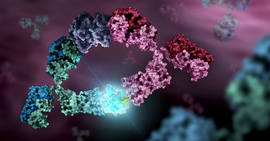
Biologic therapeutics such as monoclonal antibodies and biosimilars are complex proteins that are susceptible to post-translational modifications (PTMs). These chemical modifications can affect the performance and activity of the biologic, potentially resulting in decreased potency and increased immunogenicity. Such modifications include glycosylation, deamidation, oxidation and disulfide bond shuffling. These PTMs can be signs of protein degradation, manufacturing issues or improper storage. Several of these modifications are well characterized, and methods exist for detecting them during biologic manufacture. However, disulfide shuffling is not particularly well characterized for biologics, and no methods exist to easily detect and quantify disulfide bond shuffling in biologics.
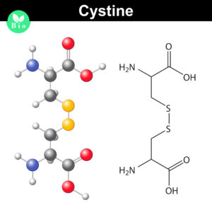
Disulfide bonds are important for protein conformation and function
Normally the cysteines in a protein will pair with a predictable or “normal” partner residue either within a polypeptide chain or between two polypeptide chains when they form disulfide bonds. These normal disulfide bonds are important for final protein conformation and stability. Indeed, disulfide bonds are considered an important quality indicator for biologics.
In a recently published study, Coghlan and colleagues designed a semi-automated method for characterizing disulfide bond shuffling on two IgG1 biologics: rituximab (originator drug Rituxan® and biosimilar Acellbia®) and bevacizumab (originator Avastin® and biosimilar Avegra®).
This metal sculpture adorns the parking garage that serves Promega Kornberg and Feynman buildings on the Madison, WI, USA campus.
World Art Day embraces art as a means for nurturing creativity and innovation, gaining greater understanding of cultural experiences that are different from our own, and showcasing the contributions of art and artists to sustainable development. It is no accident that World Art Day is celebrated on the birthday of Leonardo da Vinci, the celebrated Italian polymath who used art to express emotions, articulate technological concepts well before their time, and understand the workings of human anatomy.
Takaski Mitachi, a moderator of the 2019 global conference panel on Innovation through Art: Leveraging Disruption for a Sustainable Ecosystem, said that “Art can add value so that we could drive innovation beyond logic and data.” In a sense, the fine arts are a way of expressing our observations about the world around us, similar to how an original scientific hypothesis attempts to explain phenomena we observe and want to test. While scientific investigation builds on data and logic to test a hypothesis, art gives us a different way of knowing our world that extends beyond just data and logic. When we explore the arts, we broaden our understanding and interpretation of the world and bring these new perspectives to our scientific and technological explorations. Art is a disruptor of staid ways of thinking about and approaching problems, and like many other disruptors in our world, it can drive innovation.
In an article in the MIT Technology Review, author Sarah Lewis discusses the relationship between the arts and scientific breakthroughs. She notes one study that found a disproportionately high number of Nobel Prize-winning scientists also pursue art, writing or music as serious avocations. When asked why achievement in art and science seem to go together, she replied: “What the arts allow us to do is develop the muscle required for discernment and also strengthen our sense of agency to determine for ourselves how we’re going to tackle a given problem…Ultimately it’s up to the person creating the work to determine what that path is, and that kind of agency is what’s required for innovation.”
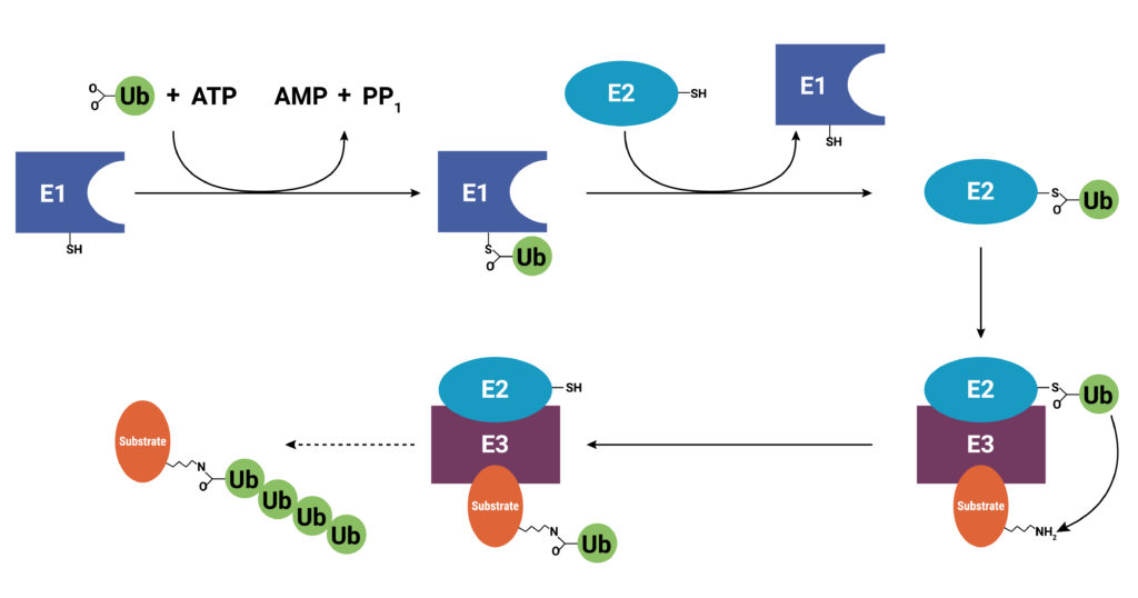
With use and time things wear out. Tires get worn on a car, and you have the old tires removed, recycled, and replaced with new ones. Sometimes a part or piece of something isn’t made properly. For instance, if you are assembling a piece of furniture and you find a screw with no threads, you throw it out and get a screw that was made properly. The same thing holds true for cells. Components wear out (like tires) or get improperly made (a screw with no threads), or they simply have a limited lifetime so that they are available in the cell only when needed. These used and worn components need to be removed from the cell. One system that allows cells to recycle components and remove old or improperly functioning proteins is the Ubiquitin-Proteasome System (UPS). The UPS system relies on a series of small peptide tags, ubiquitin, to mark a protein for degradation. Researchers are now harnessing the UPS to target aberrant proteins in diseased cells through PROteolysis TArgeting Chimeras or PROTACs. PROTACs hold promise as highly efficacious therapeutics that can be directed to eliminate only a single protein. To take full advantage of the power of PROTACs, researchers need to understand the molecular underpinnings that are responsible for successful protein degradation. Here we review a paper that seeks to develop a computer model for predicting whether PROTAC ternary complex formation leads to ubiquitination and successful degradation of a target protein.
Proteins are targeted for degradation by the proteasome. A small chain of ubiquitin peptides (Ub) is added to available lysine residues of the target protein through the actions of three enzymes: E1, ubiquitin-activating enzyme; E2, ubiquitin-conjugating enzyme; and E3 ubiquitin ligase. After the addition of the Ub chain, the proteasome is recruited and the protein degraded.
Addressing the Intractable Target
Research to understand diseases including cancers, neurodegeneration, and auto-immune conditions has revealed that in many disease states, affected cells produce growth factors or enzymes that are constitutively active (“always on”). These proteins are targets for small molecule inhibitors that bind specific sites preventing the constitutive activity or signaling. More recently, biologics, or protein-based therapeutics, including monoclonal antibodies (mAb), have been developed that can bind and block inappropriate signaling pathways, especially those that allow cancer cells to escape immune system surveillance.
Unfortunately, up to 85% of targets have proven intractable to small molecule inhibitors, or they are not suitable for a biologics approach. Oftentimes, the target protein doesn’t have a great place to bind a small molecule, so even though inhibitors might exist they cannot bind well enough to be effective. Or, as in the case of many cancers, the diseased cell manages to overcome the effect of the inhibitor by overexpressing the target. Still other aberrant proteins associated with diseases haven’t gained function to cause a disease; they have instead, lost function, so designing an inhibitor of the protein is not a workable strategy. Enter the PROTAC.
Research in animal models shows physical exercise can induce changes in the brain. In humans, studies also revealed changes in brain physiology and function resulting from physical exercise, including increased hippocampal and cognitive performance (1). Several studies in mice and rats also demonstrated that exercise can improve learning and memory and decrease neuroinflammation in models of Alzheimer’s disease and other neurodegenerative pathologies (2); these benefits are tied to increased plasticity and decreased inflammation in the hippocampus in mice (2). If regular time pounding the pavement does improve brain function, what is the underlying molecular biology of exercise-induced neuroprotection? Can we identify the cellular pathways and components involved? Can we detect important components in blood plasma? And, is the benefit of these components transferrable between organisms? De Miguel and colleagues set out to answer these questions and describe their results in a recent study published in Nature.
This summer, Dr. Anette Leue, Director of Digital Marketing and PR Promega GmbH, represented Promega Corporation in Sustainability Day activities sponsored by Smart Lab Connects. Dr. Leue presented Promega Corporation’s corporate responsibility activities and joined a panel discussion about global responsibility with representatives from Eppendorf, Max Planck Sustainability Network, and NIUB Sustainability Consultants.
As the Sustainability Day activities progressed, what became apparent is that calls for sustainable business growth are coming from all directions. Customers of life sciences companies are asking, “what are you doing to be a responsible company”? And, employees also are asking the same question of their employers. This interest sustainability and global responsibility by customers, employees and local communities is bringing into sharp focus the activities of companies to be good corporate citizens. Sustainability and global responsibility programs are no longer nice extras for life science companies, but rather are requirements for doing business.
“Sustainability is not a “nice to have”, but something that should be intrinsically implemented in the companies.”
September 11, 2001 is the day that will live in infamy for my generation. On that beautiful late summer day, I was at my desk working on the Fall issue of Neural Notes magazine when a colleague learned of the first plane hitting the World Trade Center. As the morning wore on, we learned quickly that it wasn’t just one plane, and it wasn’t just the World Trade Center.
Information was sparse. The world wide web was incredibly slow, and social media wasn’t much of a thing—nothing more than a few listservs for the life sciences. Someone managed to find a TV with a rabbit-eared, foil-covered antenna, and we gathered in the cafeteria of Promega headquarters—our shock growing as more footage became available. At Promega, conversation immediately turned to how we could bring our DNA forensic analysis expertise to help and support the authorities with the identification of victims and cataloguing of reference samples.
Just as the internet and social media have evolved into faster and more powerful means of communication—no longer do we rely on TVs with antennas for breaking news—the technology that is used to identify victims of a tragedy from partial remains like bone fragments and teeth has also evolved to be faster and more powerful.
Teeth and Bones: Then and Now
“Bones tell me the story of a person’s life—how old they were, what their gender was, their ancestral background.” Kathy Reichs
Many stories, both fact and fiction, start with a discovery of bones from a burial site or other scene. Bones can be recovered from harsh environments, having been exposed to extreme heat, time, acidic soils, swamps, chemicals, animal activities, water, or fires and explosions. These exposures degrade the sample and make recovering DNA from the cells deep within the bone matrix difficult.
XWe use cookies and similar technologies to make our website work, run analytics, improve our website, and show you personalized content and advertising. Some of these cookies are essential for our website to work. For others, we won’t set them unless you accept them. To learn more about our approach to Privacy we invite you to Read More
By clicking “Accept All”, you consent to the use of ALL the cookies. However you may visit Cookie Settings to provide a controlled consent.
We use cookies and similar technologies to make our website work, run analytics, improve our website, and show you personalized content and advertising. Some of these cookies are essential for our website to work. For others, we won’t set them unless you accept them. To find out more about cookies and how to manage cookies, read our Cookie Policy.
If you are located in the EEA, the United Kingdom, or Switzerland, you can change your settings at any time by clicking Manage Cookie Consent in the footer of our website.
Necessary cookies are absolutely essential for the website to function properly. These cookies ensure basic functionalities and security features of the website, anonymously.
Cookie
Duration
Description
cookielawinfo-checbox-analytics
11 months
This cookie is set by GDPR Cookie Consent plugin. The cookie is used to store the user consent for the cookies in the category "Analytics".
cookielawinfo-checbox-functional
11 months
The cookie is set by GDPR cookie consent to record the user consent for the cookies in the category "Functional".
cookielawinfo-checbox-others
11 months
This cookie is set by GDPR Cookie Consent plugin. The cookie is used to store the user consent for the cookies in the category "Other.
cookielawinfo-checkbox-advertisement
1 year
The cookie is set by GDPR cookie consent to record the user consent for the cookies in the category "Advertisement".
cookielawinfo-checkbox-necessary
11 months
This cookie is set by GDPR Cookie Consent plugin. The cookies is used to store the user consent for the cookies in the category "Necessary".
cookielawinfo-checkbox-performance
11 months
This cookie is set by GDPR Cookie Consent plugin. The cookie is used to store the user consent for the cookies in the category "Performance".
gdpr_status
6 months 2 days
This cookie is set by the provider Media.net. This cookie is used to check the status whether the user has accepted the cookie consent box. It also helps in not showing the cookie consent box upon re-entry to the website.
lang
This cookie is used to store the language preferences of a user to serve up content in that stored language the next time user visit the website.
viewed_cookie_policy
11 months
The cookie is set by the GDPR Cookie Consent plugin and is used to store whether or not user has consented to the use of cookies. It does not store any personal data.
Analytical cookies are used to understand how visitors interact with the website. These cookies help provide information on metrics the number of visitors, bounce rate, traffic source, etc.
Cookie
Duration
Description
SC_ANALYTICS_GLOBAL_COOKIE
10 years
This cookie is associated with Sitecore content and personalization. This cookie is used to identify the repeat visit from a single user. Sitecore will send a persistent session cookie to the web client.
vuid
2 years
This domain of this cookie is owned by Vimeo. This cookie is used by vimeo to collect tracking information. It sets a unique ID to embed videos to the website.
WMF-Last-Access
1 month 18 hours 24 minutes
This cookie is used to calculate unique devices accessing the website.
_ga
2 years
This cookie is installed by Google Analytics. The cookie is used to calculate visitor, session, campaign data and keep track of site usage for the site's analytics report. The cookies store information anonymously and assign a randomly generated number to identify unique visitors.
_gid
1 day
This cookie is installed by Google Analytics. The cookie is used to store information of how visitors use a website and helps in creating an analytics report of how the website is doing. The data collected including the number visitors, the source where they have come from, and the pages visted in an anonymous form.
Advertisement cookies are used to provide visitors with relevant ads and marketing campaigns. These cookies track visitors across websites and collect information to provide customized ads.
Cookie
Duration
Description
IDE
1 year 24 days
Used by Google DoubleClick and stores information about how the user uses the website and any other advertisement before visiting the website. This is used to present users with ads that are relevant to them according to the user profile.
test_cookie
15 minutes
This cookie is set by doubleclick.net. The purpose of the cookie is to determine if the user's browser supports cookies.
VISITOR_INFO1_LIVE
5 months 27 days
This cookie is set by Youtube. Used to track the information of the embedded YouTube videos on a website.
Performance cookies are used to understand and analyze the key performance indexes of the website which helps in delivering a better user experience for the visitors.
Cookie
Duration
Description
YSC
session
This cookies is set by Youtube and is used to track the views of embedded videos.
_gat_UA-62336821-1
1 minute
This is a pattern type cookie set by Google Analytics, where the pattern element on the name contains the unique identity number of the account or website it relates to. It appears to be a variation of the _gat cookie which is used to limit the amount of data recorded by Google on high traffic volume websites.