MISpheroID: A Knowledgebase to Improve Reproducibility in Spheroid Research

Spheroid research is  now a common component of cell biology and drug discovery science

Advantages of Spheroids

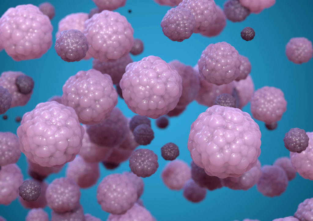
In the past decade, there has been a sharp rise in studies using spheroids as cell models for basic research and drug discovery. Spheroids are self-organized aggregation of cells that form a spherical mass, and they have become widely popular because they are much more physiologically relevant compared to flat 2D cell cultures.

In spheroids, the inner cells have less access to nutrients and oxygen compared to the outer layer, forming a natural gradient. As a result, metabolite concentration and cellular state such as proliferation and differentiation, can be very different at the periphery compared to the inner core. This phenomenon, known as “heterogeneity”, makes 3D tumor spheroids much more representative of actual tumors in the human body.

Continue reading “MISpheroID: A Knowledgebase to Improve Reproducibility in Spheroid Research”

Overcoming Challenges to Detect Apoptosis in 3D Cell Structures

This blog is written by guest author, Maggie Bach, Sr. Product Manager, Promega Corporation.

Researchers are increasingly relying on cells grown in three-dimensional (3D) structures to help answer their research questions. Monolayer, or 2D cell culture, was the go-to cell culture method for the past century. Now, the need to better represent in vivo conditions is driving the adoption of 3D cell culture models. Cells grown in 3D structures better mimic tissue-like structures, better exhibit differentiated cellular functions, and better predict in vivo responses to drug treatment.

Switching to 3D cell culture models comes with challenges. Methods to interrogate these models need to be adaptable and reliable for the many types of 3D models. Some of the most popular 3D models include spheroids grown in ultra-low attachment plates, and cells grown in an extracellular matrix, such as Matrigel® from Corning. Even more complex models include medium flow over the cells in microfluidic or organ-on-a-chip devices. Will an assay originally developed for cells grown in monolayer perform consistently with various 3D models? How is measuring a cellular marker different when cells are grown in 3D models compared to monolayer growth?

Close up of cells in 3D culture apparatus. 3D Cell Structures Provide Challenges for Measuring Markers of Cellular Activitiy
3D Cell Structures Provide Challenges for Measuring Markers of Cellular Activitiy
Continue reading “Overcoming Challenges to Detect Apoptosis in 3D Cell Structures”

Out-FOXOing High-Stage Neuroblastoma

Fluorescence microscopy of neuroblastoma cells.

In recent years, scientists have been hot on the trail of transcription factor FOXO3, tracing its involvement in various tumor-centric activities comprising the many trademarks of cancer, from drug resistance to metastasis to tumor angiogenesis.

FOXO3 is a member of the O sub-class of the forkhead box family of transcription factors. The forkhead box (FOX) family is characterized by a fork head DNA-binding domain (DBD), comprised of around 100 amino acids. They have also proven themselves to be a family of many hats, functioning in diverse roles ranging from metabolism, immunology, cell-cycle control, development, as well as cancer (1). The forkhead box O (FOXO) sub-class alone has demonstrated involvement in a variety of cellular outcomes, from drug resistance and longevity to apoptosis induction.

Due to its pro-apoptotic and anti-proliferative proclivity, FOXO3 has been previously identified as a tumor suppressor gene. However, more and more studies have begun to flip the narrative on FOXO3, portraying it more as a devoted henchman, due to its roles in drug and radiotherapy resistance, cell-cycle arrest and long-term maintenance of leukemia-initiating stem cells in a variety of cancer types, including breast cancer, pancreatic cancer, glioblastoma, and both acute and chronic myeloid leukemia.

Continue reading “Out-FOXOing High-Stage Neuroblastoma”

Improving Cancer Drug Screening with 3D Cell Culture

Differential contrast image of HCT116 colon cancer spheroid grown in a 96-well hanging-drop platform after seeding with 800 cells. Copyright Promega Corporation.
Tissue culture using primary or cultured cell lines has long been a mainstay of testing compounds for inhibiting cell growth or promoting apoptosis during screening for cancer drugs. However, the standard culture conditions result in monolayers of cells, dividing and growing across the bottom of a well, plate or flask in a single layer. The drawback of this technique is that organisms do not come in monolayers; a three-dimensional (3D) spheroid is closer to the in vivo state, especially if the spheroids are made up of more than one cell type like tumors in multicellular organisms. Even more beneficial would be using 3D cultured cells in high-throughput screening to facilitate compound profiling for target effectiveness and cytotoxicity. In a recent PLOS ONE article, researchers used normal and breast cancer cells both in monoculture and coculture to test a set of compounds and found results differed between 2D and 3D cultured cells. Continue reading “Improving Cancer Drug Screening with 3D Cell Culture”